 Movie
Movie Controller

[English] 日本語
 Yorodumi
- PDB-3vdj: Crystal structure of circumsporozoite protein aTSR domain, R32 na... -
+ Open data

- Basic information
| Entry | Database: PDB / ID: 3vdj | ||||||
|---|---|---|---|---|---|---|---|
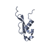
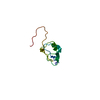
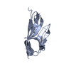
| Title | Crystal structure of circumsporozoite protein aTSR domain, R32 native form | ||||||
 Components Components | Circumsporozoite (CS) protein | ||||||
 Keywords Keywords | CELL INVASION / TSR / aTSR | ||||||
| Function / homology |  Function and homology information Function and homology informationhost cell surface binding / symbiont entry into host / entry into host cell by a symbiont-containing vacuole / heparan sulfate proteoglycan binding / side of membrane / cell surface / plasma membrane / cytoplasm Similarity search - Function | ||||||
| Biological species |  | ||||||
| Method |  X-RAY DIFFRACTION / SYNCHROTRON / MOLECULAR REPLACEMENT / Resolution: 1.698 Å X-RAY DIFFRACTION / SYNCHROTRON / MOLECULAR REPLACEMENT / Resolution: 1.698 Å | ||||||
 Authors Authors | Doud, M.B. / Koksal, A.C. / Mi, L.Z. / Song, G. / Lu, C. / Springer, T.A. | ||||||
 Citation Citation | Journal: Proc.Natl.Acad.Sci.USA / Year: 2012 Title: Unexpected fold in the circumsporozoite protein target of malaria vaccines. Authors: Doud, M.B. / Koksal, A.C. / Mi, L.Z. / Song, G. / Lu, C. / Springer, T.A. | ||||||
| History |
|
- Structure visualization
| Structure viewer | Molecule: MolmilJmol/JSmol |
|---|
- Downloads & links
-Download
| PDBx/mmCIF format | 3vdj.cif.gz | 44.9 KB | Display | PDBx/mmCIF format |
|---|---|---|---|---|
| PDB format | pdb3vdj.ent.gz | 31.7 KB | Display | PDB format |
| PDBx/mmJSON format | 3vdj.json.gz | Tree view | PDBx/mmJSON format | |
| Others |  Other downloads Other downloads |
-Validation report
| Arichive directory | https://data.pdbj.org/pub/pdb/validation_reports/vd/3vdjftp://data.pdbj.org/pub/pdb/validation_reports/vd/3vdj | HTTPS FTP |
|---|
-Related structure data
| Related structure data |  3vdkSC  3vdlC S: Starting model for refinement C: citing same article ( |
|---|---|
| Similar structure data |


-Links
 PDBj
PDBj- Assembly
| Deposited unit | 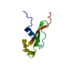
| |||||||||||||||||||||
|---|---|---|---|---|---|---|---|---|---|---|---|---|---|---|---|---|---|---|---|---|---|---|
| 1 |
| |||||||||||||||||||||
| 2 | 
| |||||||||||||||||||||
| Unit cell |
| |||||||||||||||||||||
| Components on special symmetry positions |
|
-Components
| #1: Protein | Mass: 8902.075 Da / Num. of mol.: 1 / Fragment: alpha-TSR domain (UNP residues 310-374) Source method: isolated from a genetically manipulated source Source: (gene. exp.) Strain: 3D7 / Gene: PFC0210c / Production host:  Pichia pastoris (fungus) / Strain (production host): GS115 / References: UniProt: Q7K740 Pichia pastoris (fungus) / Strain (production host): GS115 / References: UniProt: Q7K740 |
|---|---|
| #2: Water | ChemComp-HOH /  Mass: 18.015 Da / Num. of mol.: 103 / Source method: isolated from a natural source / Formula: H2O Mass: 18.015 Da / Num. of mol.: 103 / Source method: isolated from a natural source / Formula: H2O |
| Has protein modification | Y |
-Experimental details
-Experiment
| Experiment | Method: X-RAY DIFFRACTION / Number of used crystals: 1 |
|---|
- Sample preparation
| Crystal | Density Matthews: 2.06 Å3/Da / Density % sol: 40.32 % |
|---|---|
| Crystal grow | Temperature: 295 K / Method: vapor diffusion, hanging drop / pH: 4 Details: 0.1 M citrate, pH 4.0, 1 M lithium chloride, 20% PEG6000, VAPOR DIFFUSION, HANGING DROP, temperature 295K |
-Data collection
| Diffraction | Mean temperature: 93 K |
|---|---|
| Diffraction source | Source: SYNCHROTRON / Site: APS  / Beamline: 24-ID-E / Wavelength: 0.97949 Å / Beamline: 24-ID-E / Wavelength: 0.97949 Å |
| Detector | Type: ADSC QUANTUM 315 / Detector: CCD / Date: Jul 15, 2010 |
| Radiation | Monochromator: single crystal Si(220) / Protocol: SINGLE WAVELENGTH / Monochromatic (M) / Laue (L): M / Scattering type: x-ray |
| Radiation wavelength | Wavelength: 0.97949 Å / Relative weight: 1 |
| Reflection | Resolution: 1.698→50 Å / Num. all: 8234 / Num. obs: 8160 / % possible obs: 99.1 % / Observed criterion σ(I): 5 / Redundancy: 4.9 % / Biso Wilson estimate: 21.8 Å2 / Rmerge(I) obs: 0.057 / Net I/σ(I): 22.7 |
| Reflection shell | Resolution: 1.698→1.73 Å / Rmerge(I) obs: 0.351 / Mean I/σ(I) obs: 3.6 / % possible all: 91.1 |
- Processing
| Software |
| ||||||||||||||||||||||||||||||||||||||||||||||||||||||||||||||||||||||||||||||||||||||||||||||||||||||||||||||||||||||||||||||||||||||||||||||||||||||||||||||||||||||||||||||||||||||||||||||||||||||||
|---|---|---|---|---|---|---|---|---|---|---|---|---|---|---|---|---|---|---|---|---|---|---|---|---|---|---|---|---|---|---|---|---|---|---|---|---|---|---|---|---|---|---|---|---|---|---|---|---|---|---|---|---|---|---|---|---|---|---|---|---|---|---|---|---|---|---|---|---|---|---|---|---|---|---|---|---|---|---|---|---|---|---|---|---|---|---|---|---|---|---|---|---|---|---|---|---|---|---|---|---|---|---|---|---|---|---|---|---|---|---|---|---|---|---|---|---|---|---|---|---|---|---|---|---|---|---|---|---|---|---|---|---|---|---|---|---|---|---|---|---|---|---|---|---|---|---|---|---|---|---|---|---|---|---|---|---|---|---|---|---|---|---|---|---|---|---|---|---|---|---|---|---|---|---|---|---|---|---|---|---|---|---|---|---|---|---|---|---|---|---|---|---|---|---|---|---|---|---|---|---|---|
| Refinement | Method to determine structure: MOLECULAR REPLACEMENT Starting model: PDB ENTRY 3VDK Resolution: 1.698→47.774 Å / SU ML: 0.4 / σ(F): 0 / Phase error: 21.69 / Stereochemistry target values: ML
| ||||||||||||||||||||||||||||||||||||||||||||||||||||||||||||||||||||||||||||||||||||||||||||||||||||||||||||||||||||||||||||||||||||||||||||||||||||||||||||||||||||||||||||||||||||||||||||||||||||||||
| Solvent computation | Shrinkage radii: 0.83 Å / VDW probe radii: 1.1 Å / Solvent model: FLAT BULK SOLVENT MODEL / Bsol: 44.857 Å2 / ksol: 0.389 e/Å3 | ||||||||||||||||||||||||||||||||||||||||||||||||||||||||||||||||||||||||||||||||||||||||||||||||||||||||||||||||||||||||||||||||||||||||||||||||||||||||||||||||||||||||||||||||||||||||||||||||||||||||
| Displacement parameters |
| ||||||||||||||||||||||||||||||||||||||||||||||||||||||||||||||||||||||||||||||||||||||||||||||||||||||||||||||||||||||||||||||||||||||||||||||||||||||||||||||||||||||||||||||||||||||||||||||||||||||||
| Refinement step | Cycle: LAST / Resolution: 1.698→47.774 Å
| ||||||||||||||||||||||||||||||||||||||||||||||||||||||||||||||||||||||||||||||||||||||||||||||||||||||||||||||||||||||||||||||||||||||||||||||||||||||||||||||||||||||||||||||||||||||||||||||||||||||||
| Refine LS restraints |
| ||||||||||||||||||||||||||||||||||||||||||||||||||||||||||||||||||||||||||||||||||||||||||||||||||||||||||||||||||||||||||||||||||||||||||||||||||||||||||||||||||||||||||||||||||||||||||||||||||||||||
| LS refinement shell |
| ||||||||||||||||||||||||||||||||||||||||||||||||||||||||||||||||||||||||||||||||||||||||||||||||||||||||||||||||||||||||||||||||||||||||||||||||||||||||||||||||||||||||||||||||||||||||||||||||||||||||
| Refinement TLS params. | Method: refined / Refine-ID: X-RAY DIFFRACTION
| ||||||||||||||||||||||||||||||||||||||||||||||||||||||||||||||||||||||||||||||||||||||||||||||||||||||||||||||||||||||||||||||||||||||||||||||||||||||||||||||||||||||||||||||||||||||||||||||||||||||||
| Refinement TLS group |
|
