- PDB-3vd1: structure of p73 DNA binding domain tetramer modulates p73 transa... -
+
Open data
ID or keywords:
Loading...
-
Basic information
Entry
Database: PDB / ID: 3vd1
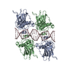
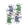
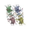
Title
structure of p73 DNA binding domain tetramer modulates p73 transactivation
Components
DNA (5'-D(*CP*GP*GP*GP*CP*AP*TP*GP*CP*CP*CP*G)-3')
Tumor protein p73
Keywords
ANTITUMOR PROTEIN/DNA / PROTEIN DNA COMPLEX / beta-immunoglobulin-like fold / TUMOUR SUPPRESSOR / ANTITUMOR PROTEIN-DNA complex
Function / homology
Function and homology information
positive regulation of lung ciliated cell differentiation / cerebrospinal fluid secretion / negative regulation of cardiac muscle cell proliferation / positive regulation of intrinsic apoptotic signaling pathway in response to DNA damage by p53 class mediator / TP53 Regulates Transcription of Death Receptors and Ligands / Activation of PUMA and translocation to mitochondria / Regulation of TP53 Activity through Association with Co-factors / negative regulation of neuron differentiation / digestive tract morphogenesis / TP53 Regulates Transcription of Caspase Activators and Caspases ...positive regulation of lung ciliated cell differentiation / cerebrospinal fluid secretion / negative regulation of cardiac muscle cell proliferation / positive regulation of intrinsic apoptotic signaling pathway in response to DNA damage by p53 class mediator / TP53 Regulates Transcription of Death Receptors and Ligands / Activation of PUMA and translocation to mitochondria / Regulation of TP53 Activity through Association with Co-factors / negative regulation of neuron differentiation / digestive tract morphogenesis / TP53 Regulates Transcription of Caspase Activators and Caspases / TP53 Regulates Transcription of Genes Involved in Cytochrome C Release / TP53 regulates transcription of several additional cell death genes whose specific roles in p53-dependent apoptosis remain uncertain / positive regulation of oligodendrocyte differentiation / intrinsic apoptotic signaling pathway in response to DNA damage by p53 class mediator / positive regulation of cell size / neuron development / mismatch repair / MDM2/MDM4 family protein binding / regulation of mitotic cell cycle / release of cytochrome c from mitochondria / transcription corepressor binding / post-embryonic development / hippocampus development / protein tetramerization / kidney development / intrinsic apoptotic signaling pathway in response to DNA damage / p53 binding / cell junction / RUNX1 regulates transcription of genes involved in differentiation of HSCs / regulation of gene expression / DNA-binding transcription activator activity, RNA polymerase II-specific / DNA-binding transcription factor binding / negative regulation of neuron apoptotic process / RNA polymerase II-specific DNA-binding transcription factor binding / DNA-binding transcription factor activity, RNA polymerase II-specific / transcription cis-regulatory region binding / regulation of cell cycle / positive regulation of MAPK cascade / ciliary basal body / RNA polymerase II cis-regulatory region sequence-specific DNA binding / response to xenobiotic stimulus / inflammatory response / DNA-binding transcription factor activity / negative regulation of cell population proliferation / intracellular membrane-bounded organelle / DNA damage response / centrosome / regulation of transcription by RNA polymerase II / protein kinase binding / chromatin / positive regulation of DNA-templated transcription / Golgi apparatus / positive regulation of transcription by RNA polymerase II / nucleoplasm / metal ion binding / identical protein binding / nucleus / plasma membrane / cytosol Similarity search - Function
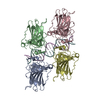
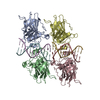
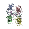
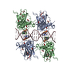
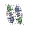
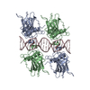
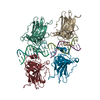
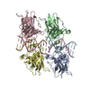
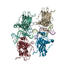
A: Tumor protein p73 B: Tumor protein p73 C: Tumor protein p73 D: Tumor protein p73 I: Tumor protein p73 J: Tumor protein p73 K: Tumor protein p73 L: Tumor protein p73 E: DNA (5'-D(*CP*GP*GP*GP*CP*AP*TP*GP*CP*CP*CP*G)-3') F: DNA (5'-D(*CP*GP*GP*GP*CP*AP*TP*GP*CP*CP*CP*G)-3') G: DNA (5'-D(*CP*GP*GP*GP*CP*AP*TP*GP*CP*CP*CP*G)-3') H: DNA (5'-D(*CP*GP*GP*GP*CP*AP*TP*GP*CP*CP*CP*G)-3') M: DNA (5'-D(*CP*GP*GP*GP*CP*AP*TP*GP*CP*CP*CP*G)-3') N: DNA (5'-D(*CP*GP*GP*GP*CP*AP*TP*GP*CP*CP*CP*G)-3') O: DNA (5'-D(*CP*GP*GP*GP*CP*AP*TP*GP*CP*CP*CP*G)-3') P: DNA (5'-D(*CP*GP*GP*GP*CP*AP*TP*GP*CP*CP*CP*G)-3') hetero molecules
A: Tumor protein p73 B: Tumor protein p73 C: Tumor protein p73 D: Tumor protein p73 E: DNA (5'-D(*CP*GP*GP*GP*CP*AP*TP*GP*CP*CP*CP*G)-3') F: DNA (5'-D(*CP*GP*GP*GP*CP*AP*TP*GP*CP*CP*CP*G)-3') G: DNA (5'-D(*CP*GP*GP*GP*CP*AP*TP*GP*CP*CP*CP*G)-3') H: DNA (5'-D(*CP*GP*GP*GP*CP*AP*TP*GP*CP*CP*CP*G)-3') hetero molecules
I: Tumor protein p73 J: Tumor protein p73 K: Tumor protein p73 L: Tumor protein p73 M: DNA (5'-D(*CP*GP*GP*GP*CP*AP*TP*GP*CP*CP*CP*G)-3') N: DNA (5'-D(*CP*GP*GP*GP*CP*AP*TP*GP*CP*CP*CP*G)-3') O: DNA (5'-D(*CP*GP*GP*GP*CP*AP*TP*GP*CP*CP*CP*G)-3') P: DNA (5'-D(*CP*GP*GP*GP*CP*AP*TP*GP*CP*CP*CP*G)-3') hetero molecules
In the structure databanks used in Yorodumi, some data are registered as the other names, "COVID-19 virus" and "2019-nCoV". Here are the details of the virus and the list of structure data.
Jan 31, 2019. EMDB accession codes are about to change! (news from PDBe EMDB page)
EMDB accession codes are about to change! (news from PDBe EMDB page)
The allocation of 4 digits for EMDB accession codes will soon come to an end. Whilst these codes will remain in use, new EMDB accession codes will include an additional digit and will expand incrementally as the available range of codes is exhausted. The current 4-digit format prefixed with “EMD-” (i.e. EMD-XXXX) will advance to a 5-digit format (i.e. EMD-XXXXX), and so on. It is currently estimated that the 4-digit codes will be depleted around Spring 2019, at which point the 5-digit format will come into force.
The EM Navigator/Yorodumi systems omit the EMD- prefix.
Related info.:Q: What is EMD? / ID/Accession-code notation in Yorodumi/EM Navigator
Yorodumi is a browser for structure data from EMDB, PDB, SASBDB, etc.
This page is also the successor to EM Navigator detail page, and also detail information page/front-end page for Omokage search.
The word "yorodu" (or yorozu) is an old Japanese word meaning "ten thousand". "mi" (miru) is to see.
Related info.:EMDB / PDB / SASBDB / Comparison of 3 databanks / Yorodumi Search / Aug 31, 2016. New EM Navigator & Yorodumi / Yorodumi Papers / Jmol/JSmol / Function and homology information / Changes in new EM Navigator and Yorodumi
 Movie
Movie Controller

 Yorodumi
 Open data
 Basic information
 Components
 Keywords
 Function and homology information
 Homo sapiens (human)
 X-RAY DIFFRACTION /
  Authors
 Citation
 Structure visualization
 Downloads & links
 Other downloads
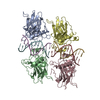


 PDBj
PDBj


 Assembly
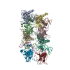


 Mass: 65.409 Da / Num. of mol.: 8 / Source method: obtained synthetically / Formula: Zn
 Mass: 18.015 Da / Num. of mol.: 188 / Source method: isolated from a natural source / Formula: H2O
 Sample preparation
 / Beamline: BL7-1 / Wavelength: 0.98
  Processing