 Movie
Movie Controller

[English] 日本語
 Yorodumi
- PDB-3v9k: Crystal structure of mouse 1-pyrroline-5-carboxylate dehydrogenas... -
+ Open data

- Basic information
| Entry | Database: PDB / ID: 3v9k | ||||||
|---|---|---|---|---|---|---|---|
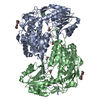
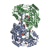
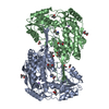
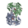
| Title | Crystal structure of mouse 1-pyrroline-5-carboxylate dehydrogenase complexed with the product glutamate | ||||||
 Components Components | Delta-1-pyrroline-5-carboxylate dehydrogenase, mitochondrial | ||||||
 Keywords Keywords | OXIDOREDUCTASE / aldehyde dehydrogenase / Rossmann fold / nucleotide binding / acting on aldehyde or oxo group of donors / NAD or NADP as acceptor / mitochondria | ||||||
| Function / homology |  Function and homology information Function and homology informationProline catabolism / Glyoxylate metabolism and glycine degradation / L-glutamate gamma-semialdehyde dehydrogenase / L-glutamate gamma-semialdehyde dehydrogenase activity / : / aldehyde dehydrogenase (NAD+) activity / mitochondrial matrix / mitochondrion / identical protein binding / cytosol Similarity search - Function | ||||||
| Biological species |  | ||||||
| Method |  X-RAY DIFFRACTION / SYNCHROTRON / MOLECULAR REPLACEMENT / Resolution: 1.501 Å X-RAY DIFFRACTION / SYNCHROTRON / MOLECULAR REPLACEMENT / Resolution: 1.501 Å | ||||||
 Authors Authors | Tanner, J.J. / Srivastava, D. | ||||||
 Citation Citation | Journal: J.Mol.Biol. / Year: 2012 Title: The Three-Dimensional Structural Basis of Type II Hyperprolinemia. Authors: Srivastava, D. / Singh, R.K. / Moxley, M.A. / Henzl, M.T. / Becker, D.F. / Tanner, J.J. | ||||||
| History |
|
- Structure visualization
| Structure viewer | Molecule: MolmilJmol/JSmol |
|---|
- Downloads & links
-Download
| PDBx/mmCIF format | 3v9k.cif.gz | 422.3 KB | Display | PDBx/mmCIF format |
|---|---|---|---|---|
| PDB format | pdb3v9k.ent.gz | 339.7 KB | Display | PDB format |
| PDBx/mmJSON format | 3v9k.json.gz | Tree view | PDBx/mmJSON format | |
| Others |  Other downloads Other downloads |
-Validation report
| Arichive directory | https://data.pdbj.org/pub/pdb/validation_reports/v9/3v9kftp://data.pdbj.org/pub/pdb/validation_reports/v9/3v9k | HTTPS FTP |
|---|
-Related structure data
| Related structure data |  3v9gC  3v9hC  3v9iC 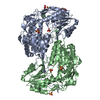 3v9jSC 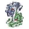 3v9lC C: citing same article ( S: Starting model for refinement |
|---|---|
| Similar structure data |


-Links
 PDBj
PDBj


- Assembly
| Deposited unit | 
| ||||||||
|---|---|---|---|---|---|---|---|---|---|
| 1 |
| ||||||||
| Unit cell |
|
-Components
| #1: Protein | Mass: 61954.215 Da / Num. of mol.: 2 / Fragment: UNP residues 21-562 Source method: isolated from a genetically manipulated source Source: (gene. exp.)  #2: Chemical |   Mass: 92.094 Da / Num. of mol.: 2 / Source method: obtained synthetically / Formula: C3H8O3 Mass: 92.094 Da / Num. of mol.: 2 / Source method: obtained synthetically / Formula: C3H8O3#3: Chemical |   Type: L-peptide linking / Mass: 147.129 Da / Num. of mol.: 2 / Source method: obtained synthetically / Formula: C5H9NO4 Type: L-peptide linking / Mass: 147.129 Da / Num. of mol.: 2 / Source method: obtained synthetically / Formula: C5H9NO4#4: Water | ChemComp-HOH / |  Mass: 18.015 Da / Num. of mol.: 896 / Source method: isolated from a natural source / Formula: H2O Mass: 18.015 Da / Num. of mol.: 896 / Source method: isolated from a natural source / Formula: H2O |
|---|
-Experimental details
-Experiment
| Experiment | Method: X-RAY DIFFRACTION / Number of used crystals: 1 |
|---|
- Sample preparation
| Crystal | Density Matthews: 2.13 Å3/Da / Density % sol: 42.15 % |
|---|---|
| Crystal grow | Temperature: 293 K / Method: vapor diffusion, sitting drop / pH: 6.5 Details: reservoir: 20-25% w/v PEG3350, 0.2 M lithium sulfate, 0.1 M Bis-Tris, pH 6.5, cryoprotectant: 25% glycerol, VAPOR DIFFUSION, SITTING DROP, temperature 293K |
-Data collection
| Diffraction | Mean temperature: 100 K | |||||||||||||||||||||||||||||||||||||||||||||||||||||||||||||||||||||||||||||
|---|---|---|---|---|---|---|---|---|---|---|---|---|---|---|---|---|---|---|---|---|---|---|---|---|---|---|---|---|---|---|---|---|---|---|---|---|---|---|---|---|---|---|---|---|---|---|---|---|---|---|---|---|---|---|---|---|---|---|---|---|---|---|---|---|---|---|---|---|---|---|---|---|---|---|---|---|---|---|
| Diffraction source | Source: SYNCHROTRON / Site: ALS  / Beamline: 4.2.2 / Wavelength: 1 Å / Beamline: 4.2.2 / Wavelength: 1 Å | |||||||||||||||||||||||||||||||||||||||||||||||||||||||||||||||||||||||||||||
| Detector | Type: NOIR-1 / Detector: CCD / Date: Apr 27, 2011 | |||||||||||||||||||||||||||||||||||||||||||||||||||||||||||||||||||||||||||||
| Radiation | Monochromator: Rosenbaum-Rock Si(111) / Protocol: SINGLE WAVELENGTH / Monochromatic (M) / Laue (L): M / Scattering type: x-ray | |||||||||||||||||||||||||||||||||||||||||||||||||||||||||||||||||||||||||||||
| Radiation wavelength | Wavelength: 1 Å / Relative weight: 1 | |||||||||||||||||||||||||||||||||||||||||||||||||||||||||||||||||||||||||||||
| Reflection | Resolution: 1.501→46.977 Å / Num. all: 168017 / Num. obs: 168017 / % possible obs: 99.6 % / Redundancy: 7.3 % / Rsym value: 0.066 / Net I/σ(I): 25.3 | |||||||||||||||||||||||||||||||||||||||||||||||||||||||||||||||||||||||||||||
| Reflection shell | Diffraction-ID: 1
|
- Processing
| Software |
| |||||||||||||||||||||||||||||||||||||||||||||||||||||||||||||||||||||||||||||
|---|---|---|---|---|---|---|---|---|---|---|---|---|---|---|---|---|---|---|---|---|---|---|---|---|---|---|---|---|---|---|---|---|---|---|---|---|---|---|---|---|---|---|---|---|---|---|---|---|---|---|---|---|---|---|---|---|---|---|---|---|---|---|---|---|---|---|---|---|---|---|---|---|---|---|---|---|---|---|
| Refinement | Method to determine structure: MOLECULAR REPLACEMENT Starting model: PDB ENTRY 3V9J Resolution: 1.501→46.977 Å / Occupancy max: 1 / Occupancy min: 0.11 / FOM work R set: 0.9229 / SU ML: 0.29 / Cross valid method: THROUGHOUT / σ(F): 0 / Phase error: 14.34 / Stereochemistry target values: ML
| |||||||||||||||||||||||||||||||||||||||||||||||||||||||||||||||||||||||||||||
| Solvent computation | Shrinkage radii: 1.06 Å / VDW probe radii: 1.3 Å / Solvent model: FLAT BULK SOLVENT MODEL / Bsol: 35.218 Å2 / ksol: 0.387 e/Å3 | |||||||||||||||||||||||||||||||||||||||||||||||||||||||||||||||||||||||||||||
| Displacement parameters | Biso max: 41.58 Å2 / Biso mean: 11.1311 Å2 / Biso min: 3.33 Å2
| |||||||||||||||||||||||||||||||||||||||||||||||||||||||||||||||||||||||||||||
| Refinement step | Cycle: LAST / Resolution: 1.501→46.977 Å
| |||||||||||||||||||||||||||||||||||||||||||||||||||||||||||||||||||||||||||||
| Refine LS restraints |
| |||||||||||||||||||||||||||||||||||||||||||||||||||||||||||||||||||||||||||||
| LS refinement shell | Refine-ID: X-RAY DIFFRACTION / Total num. of bins used: 10
|
