 Movie
Movie Controller

[English] 日本語
 Yorodumi
- PDB-3v9d: Crystal structure of the tetra-decanucleotide d(CCCCGGTACCGGGG)2 ... -
+ Open data

- Basic information
| Entry | Database: PDB / ID: 3v9d | ||||||||||||||||||
|---|---|---|---|---|---|---|---|---|---|---|---|---|---|---|---|---|---|---|---|
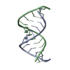
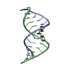
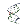
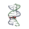
| Title | Crystal structure of the tetra-decanucleotide d(CCCCGGTACCGGGG)2 as an A-DNA duplex | ||||||||||||||||||
 Components Components | DNA (5'-D(* Keywords KeywordsDNA / Tetra-decanucleotide / A-DNA duplex / Crystal packing / A-type double helix | Function / homology | : / DNA / DNA (> 10) |  Function and homology information Function and homology informationMethod |  X-RAY DIFFRACTION / MOLECULAR REPLACEMENT / Resolution: 2.5 Å X-RAY DIFFRACTION / MOLECULAR REPLACEMENT / Resolution: 2.5 Å  Authors AuthorsMandal, P.K. / Venkadesh, S. / Gautham, N. |  CitationJournal: Acta Crystallogr.,Sect.F / Year: 2012 CitationJournal: Acta Crystallogr.,Sect.F / Year: 2012Title: Structure of the tetradecanucleotide d(CCCCGGTACCGGGG)2 as an A-DNA duplex Authors: Mandal, P.K. / Venkadesh, S. / Gautham, N. History |
|
- Structure visualization
| Structure viewer | Molecule: MolmilJmol/JSmol |
|---|
- Downloads & links
-Download
| PDBx/mmCIF format | 3v9d.cif.gz | 26.1 KB | Display | PDBx/mmCIF format |
|---|---|---|---|---|
| PDB format | pdb3v9d.ent.gz | 16.7 KB | Display | PDB format |
| PDBx/mmJSON format | 3v9d.json.gz | Tree view | PDBx/mmJSON format | |
| Others |  Other downloads Other downloads |
-Validation report
| Arichive directory | https://data.pdbj.org/pub/pdb/validation_reports/v9/3v9dftp://data.pdbj.org/pub/pdb/validation_reports/v9/3v9d | HTTPS FTP |
|---|
-Related structure data
| Related structure data | |
|---|---|
| Similar structure data |


-Links
 PDBj
PDBj


- Assembly
| Deposited unit | 
| ||||||||
|---|---|---|---|---|---|---|---|---|---|
| 1 |
| ||||||||
| Unit cell |
|
-Components
| #1: DNA chain | Mass: 4282.768 Da / Num. of mol.: 2 / Source method: obtained synthetically #2: Chemical | ChemComp-MN / |   Mass: 54.938 Da / Num. of mol.: 1 / Source method: obtained synthetically / Formula: Mn Mass: 54.938 Da / Num. of mol.: 1 / Source method: obtained synthetically / Formula: Mn#3: Water | ChemComp-HOH / |  Mass: 18.015 Da / Num. of mol.: 15 / Source method: isolated from a natural source / Formula: H2O Mass: 18.015 Da / Num. of mol.: 15 / Source method: isolated from a natural source / Formula: H2O |
|---|
-Experimental details
-Experiment
| Experiment | Method: X-RAY DIFFRACTION / Number of used crystals: 1 |
|---|
- Sample preparation
| Crystal | Density Matthews: 2.2 Å3/Da / Density % sol: 44.17 % |
|---|---|
| Crystal grow | Temperature: 293 K / Method: vapor diffusion, hanging drop / pH: 7 Details: 1mM DNA, 50mM sodium cacodylate trihydrate buffer (pH 7.0), 12mM MnCl2, 10mM spermine, equilibrated against 50 % methyl pentane diol (MPD), VAPOR DIFFUSION, HANGING DROP, temperature 293K |
-Data collection
| Diffraction | Mean temperature: 100 K |
|---|---|
| Diffraction source | Source: ROTATING ANODE / Type: BRUKER AXS MICROSTAR / Wavelength: 1.5418 Å |
| Detector | Type: MAR scanner 345 mm plate / Detector: IMAGE PLATE / Date: Jun 28, 2011 / Details: mirrors |
| Radiation | Monochromator: graphite / Protocol: SINGLE WAVELENGTH / Monochromatic (M) / Laue (L): M / Scattering type: x-ray |
| Radiation wavelength | Wavelength: 1.5418 Å / Relative weight: 1 |
| Reflection | Resolution: 2.5→29.34 Å / Num. all: 2606 / Num. obs: 2599 / % possible obs: 99.7 % / Redundancy: 4.64 % / Biso Wilson estimate: 57.3 Å2 / Rmerge(I) obs: 0.045 / Rsym value: 0.042 / Net I/σ(I): 6.6 |
| Reflection shell | Resolution: 2.5→2.59 Å / Redundancy: 4.69 % / Rmerge(I) obs: 0.282 / Mean I/σ(I) obs: 1.5 / Num. unique all: 253 / Rsym value: 0.272 / % possible all: 99.6 |
- Processing
| Software |
| |||||||||||||||||||||||||||||||||||
|---|---|---|---|---|---|---|---|---|---|---|---|---|---|---|---|---|---|---|---|---|---|---|---|---|---|---|---|---|---|---|---|---|---|---|---|---|
| Refinement | Method to determine structure: MOLECULAR REPLACEMENT Starting model: A-DNA duplex Fiber model (tetra-decanucleotide) built using Insight-II Resolution: 2.5→29.34 Å / Cor.coef. Fo:Fc: 0.96 / Cor.coef. Fo:Fc free: 0.946 / SU ML: 0.252 / Isotropic thermal model: Isotropic / Cross valid method: THROUGHOUT / ESU R: 1.252 / ESU R Free: 0.304 / Stereochemistry target values: MAXIMUM LIKELIHOOD Details: Molecular replacement was carried out using AMoRe [Navaza, 1994] of the CCP4 suite. The A-DNA duplex gave the best correlation coefficient (83%)
| |||||||||||||||||||||||||||||||||||
| Solvent computation | Ion probe radii: 0.8 Å / Shrinkage radii: 0.8 Å / VDW probe radii: 1.4 Å / Solvent model: MASK | |||||||||||||||||||||||||||||||||||
| Displacement parameters | Biso mean: 33.3799 Å2
| |||||||||||||||||||||||||||||||||||
| Refinement step | Cycle: LAST / Resolution: 2.5→29.34 Å
| |||||||||||||||||||||||||||||||||||
| Refine LS restraints |
| |||||||||||||||||||||||||||||||||||
| LS refinement shell | Resolution: 2.501→2.565 Å / Total num. of bins used: 20
|
