structural constituent of nuclear lamina / negative regulation of mesenchymal cell proliferation / protein localization to nuclear envelope / establishment or maintenance of microtubule cytoskeleton polarity / Breakdown of the nuclear lamina / ventricular cardiac muscle cell development / Depolymerization of the Nuclear Lamina / Nuclear Envelope Breakdown / nuclear pore localization / DNA double-strand break attachment to nuclear envelope ...structural constituent of nuclear lamina / negative regulation of mesenchymal cell proliferation / protein localization to nuclear envelope / establishment or maintenance of microtubule cytoskeleton polarity / Breakdown of the nuclear lamina / ventricular cardiac muscle cell development / Depolymerization of the Nuclear Lamina / Nuclear Envelope Breakdown / nuclear pore localization / DNA double-strand break attachment to nuclear envelope / lamin filament / nuclear envelope organization / XBP1(S) activates chaperone genes / nuclear lamina / Initiation of Nuclear Envelope (NE) Reformation / regulation of protein localization to nucleus / intermediate filament / regulation of telomere maintenance / negative regulation of cardiac muscle hypertrophy in response to stress / muscle organ development / nuclear migration / Deregulated CDK5 triggers multiple neurodegenerative pathways in Alzheimer's disease models / negative regulation of release of cytochrome c from mitochondria / protein localization to nucleus / regulation of cell migration / Meiotic synapsis / negative regulation of extrinsic apoptotic signaling pathway / regulation of protein stability / structural constituent of cytoskeleton / double-strand break repair via nonhomologous end joining / nuclear matrix / protein import into nucleus / cellular senescence / Signaling by BRAF and RAF1 fusions / intracellular protein localization / nuclear envelope / heterochromatin formation / site of double-strand break / nuclear membrane / cellular response to hypoxia / nuclear speck / negative regulation of cell population proliferation / positive regulation of gene expression / perinuclear region of cytoplasm / structural molecule activity / nucleoplasm / identical protein binding / nucleus / cytosol 類似検索 - 分子機能
Lamin tail domain superfamily / Lamin tail domain / Lamin Tail Domain / Lamin-tail (LTD) domain profile. / Intermediate filament protein, conserved site / Intermediate filament protein / Intermediate filament (IF) rod domain signature. / Intermediate filament, rod domain / Intermediate filament (IF) rod domain profile. / Intermediate filament protein ...Lamin tail domain superfamily / Lamin tail domain / Lamin Tail Domain / Lamin-tail (LTD) domain profile. / Intermediate filament protein, conserved site / Intermediate filament protein / Intermediate filament (IF) rod domain signature. / Intermediate filament, rod domain / Intermediate filament (IF) rod domain profile. / Intermediate filament protein / Single alpha-helices involved in coiled-coils or other helix-helix interfaces - #170 / Single alpha-helices involved in coiled-coils or other helix-helix interfaces / Up-down Bundle / Mainly Alpha 類似検索 - ドメイン・相同性
 ムービー
ムービー コントローラー


 データを開く
 基本情報
基本情報 要素
要素 キーワード
キーワード 機能・相同性情報
 Homo sapiens (ヒト)
 X線回折 /
  データ登録者
 引用
引用 構造の表示
構造の表示 ダウンロードとリンク
 その他のダウンロード


 PDBj
PDBj


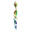
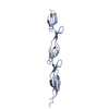
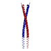
 集合体
集合体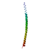

 試料調製
試料調製 / ビームライン: ID23-1 / 波長: 0.9769 Å
 解析
解析