 Movie
Movie Controller

[English] 日本語
 Yorodumi
- PDB-3uvl: Crystal structure of WDR5 in complex with the WDR5-interacting mo... -
+ Open data

- Basic information
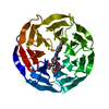
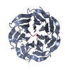
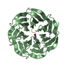
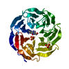
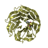
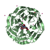
| Entry | Database: PDB / ID: 3uvl | ||||||
|---|---|---|---|---|---|---|---|
| Title | Crystal structure of WDR5 in complex with the WDR5-interacting motif of MLL3 | ||||||
 Components Components |
| ||||||
 Keywords Keywords | TRANSCRIPTION / trithorax / chromatin biology / beta-propeller / scaffolding / histone H3 / nucleus | ||||||
| Function / homology |  Function and homology information Function and homology information[histone H3]-lysine4 N-methyltransferase / histone H3K4 monomethyltransferase activity / histone H3Q5ser reader activity / histone H3K4me1 reader activity / Loss of Function of KMT2D in MLL4 Complex Formation in Kabuki Syndrome / Epigenetic regulation of gene expression by MLL3 and MLL4 complexes / MLL3/4 complex / Set1C/COMPASS complex / ATAC complex / MLL1/2 complex ...[histone H3]-lysine4 N-methyltransferase / histone H3K4 monomethyltransferase activity / histone H3Q5ser reader activity / histone H3K4me1 reader activity / Loss of Function of KMT2D in MLL4 Complex Formation in Kabuki Syndrome / Epigenetic regulation of gene expression by MLL3 and MLL4 complexes / MLL3/4 complex / Set1C/COMPASS complex / ATAC complex / MLL1/2 complex / NSL complex / histone H3K4 methyltransferase activity / Cardiogenesis / acyltransferase activity / histone methyltransferase activity / Formation of WDR5-containing histone-modifying complexes / histone methyltransferase complex / MLL1 complex / histone acetyltransferase complex / regulation of embryonic development / regulation of cell division / positive regulation of gluconeogenesis / transcription initiation-coupled chromatin remodeling / gluconeogenesis / skeletal system development / sperm principal piece / RUNX1 regulates genes involved in megakaryocyte differentiation and platelet function / PKMTs methylate histone lysines / Activation of anterior HOX genes in hindbrain development during early embryogenesis / RMTs methylate histone arginines / mitotic spindle / HATs acetylate histones / MLL4 and MLL3 complexes regulate expression of PPARG target genes in adipogenesis and hepatic steatosis / Neddylation / methylation / histone binding / transcription coactivator activity / regulation of cell cycle / regulation of transcription by RNA polymerase II / regulation of DNA-templated transcription / positive regulation of DNA-templated transcription / negative regulation of transcription by RNA polymerase II / positive regulation of transcription by RNA polymerase II / DNA binding / RNA binding / zinc ion binding / nucleoplasm / nucleus / cytosol Similarity search - Function | ||||||
| Biological species |  Homo sapiens (human) Homo sapiens (human) | ||||||
| Method |  X-RAY DIFFRACTION / SYNCHROTRON / MOLECULAR REPLACEMENT / Resolution: 2.2 Å X-RAY DIFFRACTION / SYNCHROTRON / MOLECULAR REPLACEMENT / Resolution: 2.2 Å | ||||||
 Authors Authors | Zhang, P. / Lee, H. / Brunzelle, J.S. / Couture, J.-F. | ||||||
 Citation Citation | Journal: Nucleic Acids Res. / Year: 2012 Title: The plasticity of WDR5 peptide-binding cleft enables the binding of the SET1 family of histone methyltransferases. Authors: Zhang, P. / Lee, H. / Brunzelle, J.S. / Couture, J.F. | ||||||
| History |
|
- Structure visualization
| Structure viewer | Molecule: MolmilJmol/JSmol |
|---|
- Downloads & links
-Download
| PDBx/mmCIF format | 3uvl.cif.gz | 134.3 KB | Display | PDBx/mmCIF format |
|---|---|---|---|---|
| PDB format | pdb3uvl.ent.gz | 103.1 KB | Display | PDB format |
| PDBx/mmJSON format | 3uvl.json.gz | Tree view | PDBx/mmJSON format | |
| Others |  Other downloads Other downloads |
-Validation report
| Arichive directory | https://data.pdbj.org/pub/pdb/validation_reports/uv/3uvlftp://data.pdbj.org/pub/pdb/validation_reports/uv/3uvl | HTTPS FTP |
|---|
-Related structure data
| Related structure data | 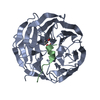 3uvkC  3uvmC  3uvnC  3uvoC  2h13S C: citing same article ( S: Starting model for refinement |
|---|---|
| Similar structure data |


-Links
 PDBj
PDBj


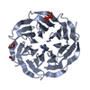
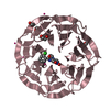
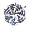
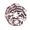
- Assembly
| Deposited unit | 
| ||||||||
|---|---|---|---|---|---|---|---|---|---|
| 1 |
| ||||||||
| Unit cell |
|
-Components
| #1: Protein | Mass: 34794.449 Da / Num. of mol.: 1 / Fragment: UNP residues 21-334 Source method: isolated from a genetically manipulated source Source: (gene. exp.) Homo sapiens (human) / Gene: WDR5, BIG3 / Production host:  |
|---|---|
| #2: Protein/peptide | Mass: 1106.254 Da / Num. of mol.: 1 / Fragment: WDR5-interacting motif (UNP residues 4707-4717) / Mutation: C4708S,S4711A / Source method: obtained synthetically / Source: (synth.) Homo sapiens (human)References: UniProt: Q8NEZ4, histone-lysine N-methyltransferase |
| #3: Water | ChemComp-HOH /  Mass: 18.015 Da / Num. of mol.: 180 / Source method: isolated from a natural source / Formula: H2O Mass: 18.015 Da / Num. of mol.: 180 / Source method: isolated from a natural source / Formula: H2O |
-Experimental details
-Experiment
| Experiment | Method: X-RAY DIFFRACTION / Number of used crystals: 1 |
|---|
- Sample preparation
| Crystal | Density Matthews: 1.98 Å3/Da / Density % sol: 37.79 % |
|---|---|
| Crystal grow | Temperature: 323 K / Method: vapor diffusion, hanging drop / pH: 8.5 Details: 0.1 M ammonium acetate, 25% PEG3350, 0.1 M Bis-Tris, pH 8.5, VAPOR DIFFUSION, HANGING DROP, temperature 323K |
-Data collection
| Diffraction | Mean temperature: 200 K |
|---|---|
| Diffraction source | Source: SYNCHROTRON / Site: APS  / Beamline: 21-ID-D / Wavelength: 1.12718 Å / Beamline: 21-ID-D / Wavelength: 1.12718 Å |
| Detector | Type: MARMOSAIC 300 mm CCD / Detector: CCD / Date: Jun 10, 2011 |
| Radiation | Monochromator: Si(111) / Protocol: SINGLE WAVELENGTH / Monochromatic (M) / Laue (L): M / Scattering type: x-ray |
| Radiation wavelength | Wavelength: 1.12718 Å / Relative weight: 1 |
| Reflection | Resolution: 2.2→54 Å / Num. all: 15066 / Num. obs: 14659 / % possible obs: 97.3 % / Observed criterion σ(F): 2 / Observed criterion σ(I): 2 / Redundancy: 7.1 % / Biso Wilson estimate: 41.96 Å2 / Rsym value: 0.075 / Net I/σ(I): 41 |
| Reflection shell | Resolution: 2.2→2.25 Å / Redundancy: 5.3 % / Mean I/σ(I) obs: 8.5 / Rsym value: 0.165 / % possible all: 81.6 |
- Processing
| Software |
| ||||||||||||||||||||||||||||||||||||||||||||||||||||||||||||||||||||||||||||||||||||||||||||||||||||||||||||
|---|---|---|---|---|---|---|---|---|---|---|---|---|---|---|---|---|---|---|---|---|---|---|---|---|---|---|---|---|---|---|---|---|---|---|---|---|---|---|---|---|---|---|---|---|---|---|---|---|---|---|---|---|---|---|---|---|---|---|---|---|---|---|---|---|---|---|---|---|---|---|---|---|---|---|---|---|---|---|---|---|---|---|---|---|---|---|---|---|---|---|---|---|---|---|---|---|---|---|---|---|---|---|---|---|---|---|---|---|---|
| Refinement | Method to determine structure: MOLECULAR REPLACEMENT Starting model: PDB ENTRY 2H13 Resolution: 2.2→40.49 Å / Cor.coef. Fo:Fc: 0.9594 / Cor.coef. Fo:Fc free: 0.9275 / Cross valid method: THROUGHOUT / σ(F): 0
| ||||||||||||||||||||||||||||||||||||||||||||||||||||||||||||||||||||||||||||||||||||||||||||||||||||||||||||
| Displacement parameters | Biso mean: 39.03 Å2
| ||||||||||||||||||||||||||||||||||||||||||||||||||||||||||||||||||||||||||||||||||||||||||||||||||||||||||||
| Refine analyze | Luzzati coordinate error obs: 0.232 Å | ||||||||||||||||||||||||||||||||||||||||||||||||||||||||||||||||||||||||||||||||||||||||||||||||||||||||||||
| Refinement step | Cycle: LAST / Resolution: 2.2→40.49 Å
| ||||||||||||||||||||||||||||||||||||||||||||||||||||||||||||||||||||||||||||||||||||||||||||||||||||||||||||
| Refine LS restraints |
| ||||||||||||||||||||||||||||||||||||||||||||||||||||||||||||||||||||||||||||||||||||||||||||||||||||||||||||
| LS refinement shell | Resolution: 2.2→2.38 Å / Total num. of bins used: 7
| ||||||||||||||||||||||||||||||||||||||||||||||||||||||||||||||||||||||||||||||||||||||||||||||||||||||||||||
| Refinement TLS params. | Method: refined / Origin x: -12.5935 Å / Origin y: 23.3525 Å / Origin z: -8.3531 Å
| ||||||||||||||||||||||||||||||||||||||||||||||||||||||||||||||||||||||||||||||||||||||||||||||||||||||||||||
| Refinement TLS group | Selection details: { A|31 - A|334 } |
