 Movie
Movie Controller

[English] 日本語
 Yorodumi
- PDB-3uus: Crystal structure of the dATP inhibited E. coli class Ia ribonucl... -
+ Open data

- Basic information
| Entry | Database: PDB / ID: 3uus | ||||||
|---|---|---|---|---|---|---|---|
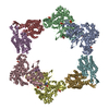
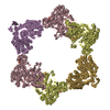
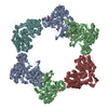
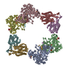
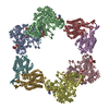
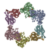
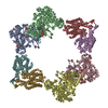
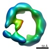
| Title | Crystal structure of the dATP inhibited E. coli class Ia ribonucleotide reductase complex | ||||||
 Components Components |
| ||||||
 Keywords Keywords | OXIDOREDUCTASE / 10 stranded alpha/beta barrel / dATP bound / di-iron | ||||||
| Function / homology |  Function and homology information Function and homology informationribonucleoside diphosphate metabolic process / 2'-deoxyribonucleotide biosynthetic process / nucleobase-containing small molecule interconversion / ribonucleoside-diphosphate reductase complex / ribonucleoside-diphosphate reductase / ribonucleoside-diphosphate reductase activity, thioredoxin disulfide as acceptor / deoxyribonucleotide biosynthetic process / protein folding chaperone / iron ion binding / ATP binding ...ribonucleoside diphosphate metabolic process / 2'-deoxyribonucleotide biosynthetic process / nucleobase-containing small molecule interconversion / ribonucleoside-diphosphate reductase complex / ribonucleoside-diphosphate reductase / ribonucleoside-diphosphate reductase activity, thioredoxin disulfide as acceptor / deoxyribonucleotide biosynthetic process / protein folding chaperone / iron ion binding / ATP binding / identical protein binding / cytoplasm / cytosol Similarity search - Function | ||||||
| Biological species |  | ||||||
| Method |  X-RAY DIFFRACTION / SYNCHROTRON / MOLECULAR REPLACEMENT / Resolution: 5.65 Å X-RAY DIFFRACTION / SYNCHROTRON / MOLECULAR REPLACEMENT / Resolution: 5.65 Å | ||||||
 Authors Authors | Zimanyi, C.M. / Drennan, C.L. | ||||||
 Citation Citation | Journal: Proc.Natl.Acad.Sci.USA / Year: 2011 Title: Structural interconversions modulate activity of Escherichia coli ribonucleotide reductase. Authors: Ando, N. / Brignole, E.J. / Zimanyi, C.M. / Funk, M.A. / Yokoyama, K. / Asturias, F.J. / Stubbe, J. / Drennan, C.L. | ||||||
| History |
|
- Structure visualization
| Structure viewer | Molecule: MolmilJmol/JSmol |
|---|
- Downloads & links
-Download
| PDBx/mmCIF format | 3uus.cif.gz | 753.5 KB | Display | PDBx/mmCIF format |
|---|---|---|---|---|
| PDB format | pdb3uus.ent.gz | 631.7 KB | Display | PDB format |
| PDBx/mmJSON format | 3uus.json.gz | Tree view | PDBx/mmJSON format | |
| Others |  Other downloads Other downloads |
-Validation report
| Arichive directory | https://data.pdbj.org/pub/pdb/validation_reports/uu/3uusftp://data.pdbj.org/pub/pdb/validation_reports/uu/3uus | HTTPS FTP |
|---|
-Related structure data


-Links
 PDBj
PDBj


- Assembly
| Deposited unit | 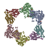
| ||||||||
|---|---|---|---|---|---|---|---|---|---|
| 1 |
| ||||||||
| Unit cell |
|
-Components
| #1: Protein | Mass: 85877.086 Da / Num. of mol.: 4 Source method: isolated from a genetically manipulated source Source: (gene. exp.) References: UniProt: P00452, ribonucleoside-diphosphate reductase #2: Protein | Mass: 43426.863 Da / Num. of mol.: 4 Source method: isolated from a genetically manipulated source Source: (gene. exp.) References: UniProt: P69924, ribonucleoside-diphosphate reductase #3: Chemical | ChemComp-DTP /   Mass: 491.182 Da / Num. of mol.: 8 / Source method: obtained synthetically / Formula: C10H16N5O12P3 Mass: 491.182 Da / Num. of mol.: 8 / Source method: obtained synthetically / Formula: C10H16N5O12P3#4: Chemical | ChemComp-FE /   Mass: 55.845 Da / Num. of mol.: 8 / Source method: obtained synthetically / Formula: Fe Mass: 55.845 Da / Num. of mol.: 8 / Source method: obtained synthetically / Formula: FeHas protein modification | Y | |
|---|
-Experimental details
-Experiment
| Experiment | Method: X-RAY DIFFRACTION / Number of used crystals: 1 |
|---|
- Sample preparation
| Crystal | Density Matthews: 3.13 Å3/Da / Density % sol: 60.7 % |
|---|---|
| Crystal grow | Temperature: 298 K / Method: vapor diffusion, hanging drop / pH: 7.5 Details: 10.8% PEG 3350, 0.18M magnesium acetate, 0.09M MOPS pH 7.5, 0.01M iron (III) chloride, 4.5% glycerol , VAPOR DIFFUSION, HANGING DROP, temperature 298K |
-Data collection
| Diffraction | Mean temperature: 100 K |
|---|---|
| Diffraction source | Source: SYNCHROTRON / Site: ALS  / Beamline: 8.2.2 / Wavelength: 0.9794 Å / Beamline: 8.2.2 / Wavelength: 0.9794 Å |
| Detector | Type: ADSC QUANTUM 315 / Detector: CCD / Date: Sep 29, 2010 |
| Radiation | Monochromator: Double crystal, Si (111) / Protocol: SINGLE WAVELENGTH / Monochromatic (M) / Laue (L): M / Scattering type: x-ray |
| Radiation wavelength | Wavelength: 0.9794 Å / Relative weight: 1 |
| Reflection | Resolution: 5.65→50 Å / Num. all: 19267 / Num. obs: 19134 / % possible obs: 99.4 % / Observed criterion σ(F): 0 / Observed criterion σ(I): 0 / Redundancy: 3.1 % / Rsym value: 0.074 / Net I/σ(I): 9.3 |
| Reflection shell | Resolution: 5.65→5.85 Å / Redundancy: 3.1 % / Mean I/σ(I) obs: 2.1 / Num. unique all: 1871 / Rsym value: 0.599 / % possible all: 99.4 |
- Processing
| Software |
| |||||||||||||||||||||||||
|---|---|---|---|---|---|---|---|---|---|---|---|---|---|---|---|---|---|---|---|---|---|---|---|---|---|---|
| Refinement | Method to determine structure: MOLECULAR REPLACEMENT Starting model: PDB entry 2R1R and PDB entry 1MXR Resolution: 5.65→50 Å / Cross valid method: THROUGHOUT / σ(F): 0 / Stereochemistry target values: Engh & Huber
| |||||||||||||||||||||||||
| Refinement step | Cycle: LAST / Resolution: 5.65→50 Å
| |||||||||||||||||||||||||
| Refine LS restraints |
|
