- PDB-3urz: Crystal structure of a putative protein binding protein (BACOVA_0... -
+
Open data
ID or keywords:
Loading...
-
Basic information
Entry
Database: PDB / ID: 3urz
Title
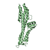
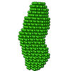
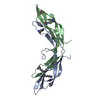
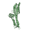
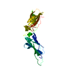
Crystal structure of a putative protein binding protein (BACOVA_03105) from Bacteroides ovatus ATCC 8483 at 2.19 A resolution
Components
Uncharacterized protein
Keywords
PROTEIN BINDING / TETRATRICOPEPTIDE REPEATS (TPR) CONTAINING PROTEIN / Structural Genomics / Joint Center for Structural Genomics / JCSG / Protein Structure Initiative / PSI-BIOLOGY
Mass: 18.015 Da / Num. of mol.: 55 / Source method: isolated from a natural source / Formula: H2O
Sequence details
THIS CONSTRUCT WAS EXPRESSED WITH A PURIFICATION TAG MGSDKIHHHHHHENLYFQG. THE TAG WAS REMOVED WITH ...THIS CONSTRUCT WAS EXPRESSED WITH A PURIFICATION TAG MGSDKIHHHHHHENLYFQG. THE TAG WAS REMOVED WITH TEV PROTEASE LEAVING ONLY A GLYCINE (0) FOLLOWED BY RESIDUES 21-227 OF THE TARGET SEQUENCE.
-
Experimental details
-
Experiment
Experiment
Method: X-RAY DIFFRACTION / Number of used crystals: 1
-
Sample preparation
Crystal
Density Matthews: 3.08 Å3/Da / Density % sol: 60.12 %
Crystal grow
Temperature: 277 K / Method: vapor diffusion, sitting drop / pH: 5.93 Details: 0.20 M zinc acetate, 16.40 % polyethylene glycol 8000, 0.1M MES pH 5.93, NANODROP, VAPOR DIFFUSION, SITTING DROP, temperature 277K
Type: MARMOSAIC 325 mm CCD / Detector: CCD / Date: Nov 22, 2010 Details: Flat mirror (vertical focusing); single crystal Si(111) bent monochromator (ho rizontal focusing)
Radiation
Monochromator: single crystal Si(111) bent / Protocol: MAD / Monochromatic (M) / Laue (L): M / Scattering type: x-ray
Radiation wavelength
ID
Wavelength (Å)
Relative weight
1
0.91837
1
2
0.98021
1
3
0.97963
1
Reflection
Resolution: 2.19→29.581 Å / Num. obs: 29857 / % possible obs: 93.3 % / Observed criterion σ(I): -3 / Biso Wilson estimate: 56.56 Å2 / Rmerge(I) obs: 0.035 / Net I/σ(I): 10.96
Reflection shell
Resolution (Å)
Rmerge(I) obs
Mean I/σ(I) obs
Num. measured obs
Num. unique obs
Diffraction-ID
% possible all
2.21-2.29
0.451
2.1
10767
5529
1
94.2
2.29-2.38
0.396
2.5
10973
5661
1
96.2
2.38-2.49
0.283
3.4
11013
5683
1
95.4
2.49-2.62
0.215
4.4
11068
5639
1
95.9
2.62-2.78
0.157
5.8
11039
5587
1
95.4
2.78-3
0.102
8.3
11123
5717
1
95.1
3-3.3
0.064
12
10435
5415
1
92.7
3.3-3.77
0.035
18.9
10465
5304
1
89.4
3.77-4.74
0.025
26
10221
5232
1
88.7
4.74-29.581
0.022
28.2
10560
5413
1
89.8
-
Phasing
Phasing
Method: MAD
-
Processing
Software
Name
Version
Classification
NB
MolProbity
3beta29
modelbuilding
PDB_EXTRACT
3.1
dataextraction
SHELX
phasing
SHARP
phasing
XSCALE
May10, 2010
datascaling
REFMAC
5.6.0117
refinement
XDS
datareduction
SHELXD
phasing
Refinement
Method to determine structure: MAD / Resolution: 2.19→29.581 Å / Cor.coef. Fo:Fc: 0.956 / Cor.coef. Fo:Fc free: 0.946 / Occupancy max: 1 / Occupancy min: 0.5 / SU B: 11.778 / SU ML: 0.158 / Cross valid method: THROUGHOUT / σ(F): 0 / ESU R: 0.235 / ESU R Free: 0.187 Stereochemistry target values: MAXIMUM LIKELIHOOD WITH PHASES Details: 1. HYDROGENS HAVE BEEN ADDED IN THE RIDING POSITIONS. 2. ATOM RECORD CONTAINS SUM OF TLS AND RESIDUAL B FACTORS. 3. ANISOU RECORD CONTAINS SUM OF TLS AND RESIDUAL U FACTORS. 4. WATERS WERE ...Details: 1. HYDROGENS HAVE BEEN ADDED IN THE RIDING POSITIONS. 2. ATOM RECORD CONTAINS SUM OF TLS AND RESIDUAL B FACTORS. 3. ANISOU RECORD CONTAINS SUM OF TLS AND RESIDUAL U FACTORS. 4. WATERS WERE EXCLUDED FROM AUTOMATIC TLS ASSIGNMENT. 5. A MET-INHIBITION PROTOCOL WAS USED FOR SELENOMETHIONINE INCORPORATION DURING PROTEIN EXPRESSION. THE OCCUPANCY OF THE SE ATOMS IN THE MSE RESIDUES WAS REDUCED TO 0.75 FOR THE REDUCED SCATTERING POWER DUE TO PARTIAL S-MET INCORPORATION. 6. PRESENCE OF ZINC IN CRYSTALLIZATION CONDITION AND ANOMALOUS DIFFERENCE FOURIERS SUPPORT THE MODELING OF ZINC (ZN) IONS. 7. PEG FRAGMENT (PG4) AND 1,2-ETHANEDIOL (EDO) MOLECULES FROM THE CRYSTALLIZATION/CRYOPROTECTION SOLUTION ARE MODELED.
Rfactor
Num. reflection
% reflection
Selection details
Rfree
0.2397
1508
5.1 %
RANDOM
Rwork
0.2173
-
-
-
obs
0.2184
29857
95.57 %
-
Solvent computation
Ion probe radii: 0.8 Å / Shrinkage radii: 0.8 Å / VDW probe radii: 1.2 Å / Solvent model: BABINET MODEL WITH MASK
In the structure databanks used in Yorodumi, some data are registered as the other names, "COVID-19 virus" and "2019-nCoV". Here are the details of the virus and the list of structure data.
Jan 31, 2019. EMDB accession codes are about to change! (news from PDBe EMDB page)
EMDB accession codes are about to change! (news from PDBe EMDB page)
The allocation of 4 digits for EMDB accession codes will soon come to an end. Whilst these codes will remain in use, new EMDB accession codes will include an additional digit and will expand incrementally as the available range of codes is exhausted. The current 4-digit format prefixed with “EMD-” (i.e. EMD-XXXX) will advance to a 5-digit format (i.e. EMD-XXXXX), and so on. It is currently estimated that the 4-digit codes will be depleted around Spring 2019, at which point the 5-digit format will come into force.
The EM Navigator/Yorodumi systems omit the EMD- prefix.
Related info.:Q: What is EMD? / ID/Accession-code notation in Yorodumi/EM Navigator
Yorodumi is a browser for structure data from EMDB, PDB, SASBDB, etc.
This page is also the successor to EM Navigator detail page, and also detail information page/front-end page for Omokage search.
The word "yorodu" (or yorozu) is an old Japanese word meaning "ten thousand". "mi" (miru) is to see.
Related info.:EMDB / PDB / SASBDB / Comparison of 3 databanks / Yorodumi Search / Aug 31, 2016. New EM Navigator & Yorodumi / Yorodumi Papers / Jmol/JSmol / Function and homology information / Changes in new EM Navigator and Yorodumi
 Movie
Movie Controller

 Yorodumi
 Open data
 Basic information
 Components
 Keywords
 Function and homology information
 Bacteroides ovatus (bacteria)
 X-RAY DIFFRACTION /
  Authors
 Citation
 Structure visualization
 Downloads & links
 Other downloads


 PDBj
PDBj


 Assembly

 Mass: 65.409 Da / Num. of mol.: 4 / Source method: obtained synthetically / Formula: Zn
 Mass: 194.226 Da / Num. of mol.: 1 / Source method: obtained synthetically / Formula: C8H18O5 / Comment: precipitant*YM
 Mass: 62.068 Da / Num. of mol.: 1 / Source method: obtained synthetically / Formula: C2H6O2
 Mass: 18.015 Da / Num. of mol.: 55 / Source method: isolated from a natural source / Formula: H2O
 Sample preparation
 / Beamline: BL11-1 / Wavelength: 0.91837,0.98021,0.97963
 Processing