[English] 日本語
 Yorodumi
- PDB-3ulm: X-ray Diffraction Studies of Ring Crystals obtained for d(CACGCG)... -
+ Open data
- Basic information
| Entry | Database: PDB / ID: 3ulm | ||||||
|---|---|---|---|---|---|---|---|
| Title | X-ray Diffraction Studies of Ring Crystals obtained for d(CACGCG).d(CGCGTG): Stage (ii) Hexagonal plates with spots | ||||||
 Components Components | 6-mer DNA | ||||||
 Keywords Keywords | DNA / Z-type DNA double helix | ||||||
| Function / homology | DNA Function and homology information Function and homology information | ||||||
| Method |  X-RAY DIFFRACTION / X-RAY DIFFRACTION /  SYNCHROTRON / SYNCHROTRON /  MOLECULAR REPLACEMENT / Resolution: 3.01 Å MOLECULAR REPLACEMENT / Resolution: 3.01 Å | ||||||
 Authors Authors | Mandal, P.K. / Venkadesh, S. / Gautham, N. | ||||||
 Citation Citation |  Journal: J.Cryst.Growth / Year: 2012 Journal: J.Cryst.Growth / Year: 2012Title: Ring crystals of oligonucleotides: Growth stages and X-ray diffraction studies Authors: Mandal, P.K. / Chandrasekaran, A.R. / Madhanagopal, B.R. / Venkadesh, S. / Gautham, N. | ||||||
| History |
|
- Structure visualization
| Structure viewer | Molecule:  Molmil Molmil Jmol/JSmol Jmol/JSmol |
|---|
- Downloads & links
- Download
| PDBx/mmCIF format |  3ulm.cif.gz 3ulm.cif.gz | 10.6 KB | Display |  PDBx/mmCIF format PDBx/mmCIF format |
|---|---|---|---|---|
| PDB format |  pdb3ulm.ent.gz pdb3ulm.ent.gz | 6.6 KB | Display |  PDB format PDB format |
| PDBx/mmJSON format |  3ulm.json.gz 3ulm.json.gz | Tree view |  PDBx/mmJSON format PDBx/mmJSON format | |
| Others |  Other downloads Other downloads |
-Validation report
| Summary document |  3ulm_validation.pdf.gz 3ulm_validation.pdf.gz | 340.3 KB | Display |  wwPDB validaton report wwPDB validaton report |
|---|---|---|---|---|
| Full document |  3ulm_full_validation.pdf.gz 3ulm_full_validation.pdf.gz | 341.9 KB | Display | |
| Data in XML |  3ulm_validation.xml.gz 3ulm_validation.xml.gz | 2.1 KB | Display | |
| Data in CIF |  3ulm_validation.cif.gz 3ulm_validation.cif.gz | 2.4 KB | Display | |
| Arichive directory |  https://data.pdbj.org/pub/pdb/validation_reports/ul/3ulm https://data.pdbj.org/pub/pdb/validation_reports/ul/3ulm ftp://data.pdbj.org/pub/pdb/validation_reports/ul/3ulm ftp://data.pdbj.org/pub/pdb/validation_reports/ul/3ulm | HTTPS FTP |
-Related structure data
- Links
Links
- Assembly
| Deposited unit | 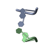
| ||||||||
|---|---|---|---|---|---|---|---|---|---|
| 1 |
| ||||||||
| Unit cell |
|
- Components
| #1: DNA chain | Mass: 1221.840 Da / Num. of mol.: 2 / Source method: obtained synthetically / Details: chemically synthesized by M/s Microsynth #2: Water | ChemComp-HOH / | Sequence details | THE ACTUAL DNA SEQUENCE FOR THE THIS STUDY IS D(CACGCG).(CGCGTG) | |
|---|
-Experimental details
-Experiment
| Experiment | Method:  X-RAY DIFFRACTION / Number of used crystals: 1 X-RAY DIFFRACTION / Number of used crystals: 1 |
|---|
- Sample preparation
| Crystal grow | Temperature: 293 K / Method: vapor diffusion, hanging drop / pH: 7 Details: 1mM DNA, 75mM sodium cacodylate trihydrate buffer (pH 7.0), 0.5mM cobalt hexammine chloride, 0.75mM spermine, equilibrated against 50% methyl pentane diol (MPD) , VAPOR DIFFUSION, HANGING DROP, temperature 293K |
|---|
-Data collection
| Diffraction | Mean temperature: 100 K |
|---|---|
| Diffraction source | Source:  SYNCHROTRON / Site: SYNCHROTRON / Site:  ELETTRA ELETTRA  / Beamline: 5.2R / Wavelength: 1.54 Å / Beamline: 5.2R / Wavelength: 1.54 Å |
| Detector | Type: MAR CCD 165 mm / Detector: CCD / Date: Jun 26, 2009 Details: The optics consist in a vertical collimating mirror, a double-crystal Si(111) monochromator followed by a toroidal bendable focussing mirror. |
| Radiation | Monochromator: a double-crystal Si(111) monochromator / Protocol: SINGLE WAVELENGTH / Monochromatic (M) / Laue (L): M / Scattering type: x-ray |
| Radiation wavelength | Wavelength: 1.54 Å / Relative weight: 1 |
| Reflection | Resolution: 3→20 Å / Num. all: 147 / Num. obs: 147 / % possible obs: 100 % / Observed criterion σ(I): 3 / Redundancy: 6.8 % / Biso Wilson estimate: 57.5 Å2 / Rmerge(I) obs: 0.111 / Rsym value: 0.0947 / Net I/σ(I): 3.3 |
| Reflection shell | Resolution: 3.01→3.12 Å / Redundancy: 6.67 % / Rmerge(I) obs: 0.404 / Mean I/σ(I) obs: 1 / Num. unique all: 12 / Rsym value: 0.352 / % possible all: 100 |
- Processing
| Software |
| |||||||||||||||||||||||||||||||||||
|---|---|---|---|---|---|---|---|---|---|---|---|---|---|---|---|---|---|---|---|---|---|---|---|---|---|---|---|---|---|---|---|---|---|---|---|---|
| Refinement | Method to determine structure:  MOLECULAR REPLACEMENT MOLECULAR REPLACEMENTStarting model: Z-type DNA Dinucleotide step built using InsightII Resolution: 3.01→15.15 Å / Cor.coef. Fo:Fc: 0.963 / Cor.coef. Fo:Fc free: 0.835 / SU B: 16.392 / SU ML: 0.296 / Cross valid method: THROUGHOUT / ESU R Free: 0.563 / Stereochemistry target values: MAXIMUM LIKELIHOOD Details: THE DNA OLIGONUCLEOTIDE HAS SIX BASE PAIRS (D(CACGCG).D(CGCGTG)) AND FORMS THE Z-TYPE DOUBLE HELICAL STRUCTURE. THE STRUCTURE HAS STATISTICAL DIS-ORDER AND COMPRISES OF A DINUCLEOTIDE STEP ...Details: THE DNA OLIGONUCLEOTIDE HAS SIX BASE PAIRS (D(CACGCG).D(CGCGTG)) AND FORMS THE Z-TYPE DOUBLE HELICAL STRUCTURE. THE STRUCTURE HAS STATISTICAL DIS-ORDER AND COMPRISES OF A DINUCLEOTIDE STEP IN THE CRYSTALLOGRAPHIC ASYMMETRIC UNIT. THE DINUCLEOTIDE STEP COULD STAND FOR EITHER CPG/CPG OR CPA/TPG. DUE TO DISORDER, THE DINUCLEOTIDE STEP WAS CONSTRUCTED AS TPG/TPG WHERE THE C5 METHYL GROUP OF THYMINE WAS ASSIGNED OCCUPANCY OF 1/6 AND N2 OF GUANINE WAS ASSIGNED OCCUPANCY OF 5/6.
| |||||||||||||||||||||||||||||||||||
| Solvent computation | Ion probe radii: 0.8 Å / Shrinkage radii: 0.8 Å / VDW probe radii: 1.4 Å / Solvent model: MASK | |||||||||||||||||||||||||||||||||||
| Displacement parameters | Biso mean: 23.785 Å2
| |||||||||||||||||||||||||||||||||||
| Refinement step | Cycle: LAST / Resolution: 3.01→15.15 Å /
| |||||||||||||||||||||||||||||||||||
| Refine LS restraints |
| |||||||||||||||||||||||||||||||||||
| LS refinement shell | Resolution: 3.013→3.088 Å / Rfactor Rfree error: 0 / Total num. of bins used: 20
|
 Movie
Movie Controller



 PDBj
PDBj


