 Movie
Movie Controller

[English] 日本語
 Yorodumi
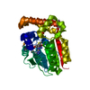
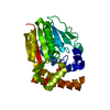
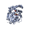
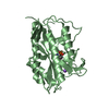
- PDB-3ujc: Phosphoethanolamine methyltransferase mutant (H132A) from Plasmod... -
+ Open data

- Basic information
| Entry | Database: PDB / ID: 3ujc | ||||||
|---|---|---|---|---|---|---|---|
| Title | Phosphoethanolamine methyltransferase mutant (H132A) from Plasmodium falciparum in complex with phosphocholine | ||||||
 Components Components | Phosphoethanolamine N-methyltransferase | ||||||
 Keywords Keywords | TRANSFERASE / Plasmodium / parasite / methyltransferase | ||||||
| Function / homology |  Function and homology information Function and homology informationphosphoethanolamine N-methyltransferase activity / phosphoethanolamine N-methyltransferase / phosphatidylcholine biosynthetic process / methylation / Golgi membrane / Golgi apparatus Similarity search - Function | ||||||
| Biological species |  | ||||||
| Method |  X-RAY DIFFRACTION / SYNCHROTRON / MOLECULAR REPLACEMENT / Resolution: 1.19 Å X-RAY DIFFRACTION / SYNCHROTRON / MOLECULAR REPLACEMENT / Resolution: 1.19 Å | ||||||
 Authors Authors | Lee, S.G. / Kim, Y. / Alpert, T.D. / Nagata, A. / Jez, J.M. | ||||||
 Citation Citation | Journal: J.Biol.Chem. / Year: 2012 Title: Structure and reaction mechanism of phosphoethanolamine methyltransferase from the malaria parasite Plasmodium falciparum: an antiparasitic drug target. Authors: Lee, S.G. / Kim, Y. / Alpert, T.D. / Nagata, A. / Jez, J.M. | ||||||
| History |
|
- Structure visualization
| Structure viewer | Molecule: MolmilJmol/JSmol |
|---|
- Downloads & links
-Download
| PDBx/mmCIF format | 3ujc.cif.gz | 130.8 KB | Display | PDBx/mmCIF format |
|---|---|---|---|---|
| PDB format | pdb3ujc.ent.gz | 101.6 KB | Display | PDB format |
| PDBx/mmJSON format | 3ujc.json.gz | Tree view | PDBx/mmJSON format | |
| Others |  Other downloads Other downloads |
-Validation report
| Arichive directory | https://data.pdbj.org/pub/pdb/validation_reports/uj/3ujcftp://data.pdbj.org/pub/pdb/validation_reports/uj/3ujc | HTTPS FTP |
|---|
-Related structure data
| Related structure data |  3uj6C  3uj7C  3uj8C  3uj9C  3ujaC  3ujbC  3ujdC C: citing same article ( |
|---|---|
| Similar structure data |


-Links
 PDBj
PDBj- Assembly
| Deposited unit | 
| ||||||||
|---|---|---|---|---|---|---|---|---|---|
| 1 |
| ||||||||
| Unit cell |
| ||||||||
| Components on special symmetry positions |
|
-Components
| #1: Protein | Mass: 31017.051 Da / Num. of mol.: 1 Source method: isolated from a genetically manipulated source Source: (gene. exp.) Gene: PMT / Production host:  |
|---|---|
| #2: Chemical | ChemComp-PC / 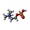  Mass: 184.151 Da / Num. of mol.: 1 / Source method: obtained synthetically / Formula: C5H15NO4P Mass: 184.151 Da / Num. of mol.: 1 / Source method: obtained synthetically / Formula: C5H15NO4P |
| #3: Water | ChemComp-HOH /  Mass: 18.015 Da / Num. of mol.: 498 / Source method: isolated from a natural source / Formula: H2O Mass: 18.015 Da / Num. of mol.: 498 / Source method: isolated from a natural source / Formula: H2O |
-Experimental details
-Experiment
| Experiment | Method: X-RAY DIFFRACTION / Number of used crystals: 1 |
|---|
- Sample preparation
| Crystal | Density Matthews: 2.32 Å3/Da / Density % sol: 46.92 % |
|---|---|
| Crystal grow | Temperature: 277 K / Method: vapor diffusion, hanging drop / pH: 6.5 Details: 20% PEG-8000, 0.1 M sodium cacodylate, pH 6.5, 0.2 M sodium acetate, 5 mM 2-mercaptoethanol, VAPOR DIFFUSION, HANGING DROP, temperature 277K |
-Data collection
| Diffraction | Mean temperature: 100 K |
|---|---|
| Diffraction source | Source: SYNCHROTRON / Site: APS  / Beamline: 19-ID / Wavelength: 0.979 Å / Beamline: 19-ID / Wavelength: 0.979 Å |
| Detector | Type: ADSC QUANTUM 315r / Detector: CCD / Date: Jul 8, 2011 |
| Radiation | Monochromator: Rosenbaum-Rock high-resolution double-crystal monochromator Protocol: SINGLE WAVELENGTH / Monochromatic (M) / Laue (L): M / Scattering type: x-ray |
| Radiation wavelength | Wavelength: 0.979 Å / Relative weight: 1 |
| Reflection | Resolution: 1.19→28.91 Å / Num. obs: 90748 / % possible obs: 99.6 % / Observed criterion σ(F): 0 / Observed criterion σ(I): 0 / Redundancy: 3.5 % / Rsym value: 0.047 / Net I/σ(I): 27.4 |
| Reflection shell | Resolution: 1.19→1.21 Å / Mean I/σ(I) obs: 2 / Rsym value: 0.409 / % possible all: 95.2 |
- Processing
| Software |
| |||||||||||||||||||||||||||||||||||||||||||||||||||||||||||||||||||||||||||||
|---|---|---|---|---|---|---|---|---|---|---|---|---|---|---|---|---|---|---|---|---|---|---|---|---|---|---|---|---|---|---|---|---|---|---|---|---|---|---|---|---|---|---|---|---|---|---|---|---|---|---|---|---|---|---|---|---|---|---|---|---|---|---|---|---|---|---|---|---|---|---|---|---|---|---|---|---|---|---|
| Refinement | Method to determine structure: MOLECULAR REPLACEMENT / Resolution: 1.19→28.91 Å / SU ML: 0.15 / σ(F): 1.35 / Phase error: 13.77 / Stereochemistry target values: ML
| |||||||||||||||||||||||||||||||||||||||||||||||||||||||||||||||||||||||||||||
| Solvent computation | Shrinkage radii: 0.83 Å / VDW probe radii: 1.1 Å / Solvent model: FLAT BULK SOLVENT MODEL / Bsol: 45.598 Å2 / ksol: 0.39 e/Å3 | |||||||||||||||||||||||||||||||||||||||||||||||||||||||||||||||||||||||||||||
| Displacement parameters |
| |||||||||||||||||||||||||||||||||||||||||||||||||||||||||||||||||||||||||||||
| Refinement step | Cycle: LAST / Resolution: 1.19→28.91 Å
| |||||||||||||||||||||||||||||||||||||||||||||||||||||||||||||||||||||||||||||
| Refine LS restraints |
| |||||||||||||||||||||||||||||||||||||||||||||||||||||||||||||||||||||||||||||
| LS refinement shell |
|
