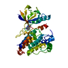
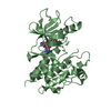
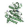
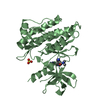
Entry Database : PDB / ID : 3ue4Title Structural and spectroscopic analysis of the kinase inhibitor bosutinib binding to the Abl tyrosine kinase domain Tyrosine-protein kinase ABL1 Keywords / / Function / homology Function Domain/homology Component
/ / / / / / / / / / / / / / / / / / / / / / / / / / / / / / / / / / / / / / / / / / / / / / / / / / / / / / / / / / / / / / / / / / / / / / / / / / / / / / / / / / / / / / / / / / / / / / / / / / / / / / / / / / / / / / / / / / / / / / / / / / / / / / / / / / / / / / / / / / / / / / / / / / / / / / Biological species Homo sapiens (human)Method / / / Resolution : 2.424 Å Authors Boxer, S.G. / Levinson, N.M. Journal : Plos One / Year : 2012Title : Structural and spectroscopic analysis of the kinase inhibitor bosutinib and an isomer of bosutinib binding to the abl tyrosine kinase domain.Authors : Levinson, N.M. / Boxer, S.G. History Deposition Oct 28, 2011 Deposition site / Processing site Revision 1.0 Apr 18, 2012 Provider / Type Revision 1.1 Apr 25, 2012 Group Revision 1.2 Feb 28, 2024 Group / Database references / Derived calculationsCategory chem_comp_atom / chem_comp_bond ... chem_comp_atom / chem_comp_bond / database_2 / struct_ref_seq_dif / struct_site Item _database_2.pdbx_DOI / _database_2.pdbx_database_accession ... _database_2.pdbx_DOI / _database_2.pdbx_database_accession / _struct_ref_seq_dif.details / _struct_site.pdbx_auth_asym_id / _struct_site.pdbx_auth_comp_id / _struct_site.pdbx_auth_seq_id
Show all Show less
 Movie
Movie Controller

 Yorodumi
 Open data
 Basic information
 Components
 Keywords
 Function and homology information
 Homo sapiens (human)
 X-RAY DIFFRACTION /
  Authors
 Citation
 Structure visualization
 Downloads & links
 Other downloads
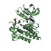


 PDBj
PDBj


 Assembly
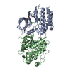


 Mass: 530.446 Da / Num. of mol.: 2 / Source method: obtained synthetically / Formula: C26H29Cl2N5O3 / Comment: inhibitor, medication*YM
 Mass: 18.015 Da / Num. of mol.: 152 / Source method: isolated from a natural source / Formula: H2O
 Sample preparation
 / Beamline: BL12-2 / Wavelength: 0.9795 Å
 Processing