 Movie
Movie Controller

[English] 日本語
 Yorodumi
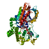
- PDB-3ucl: Cyclohexanone-bound crystal structure of cyclohexanone monooxygen... -
+ Open data

- Basic information
| Entry | Database: PDB / ID: 3ucl | ||||||
|---|---|---|---|---|---|---|---|
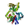
| Title | Cyclohexanone-bound crystal structure of cyclohexanone monooxygenase in the Rotated conformation | ||||||
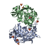
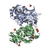
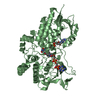
 Components Components | Cyclohexanone monooxygenase | ||||||
 Keywords Keywords | OXIDOREDUCTASE / Baeyer-Villiger monooxygenase / Baeyer-Villiger oxidation / biocatalysis / flavoprotein / green chemistry / protein engineering / Rossmann fold / FAD / NADPH / cyclohexanone / oxygen / Cytosolic (bacterial) | ||||||
| Function / homology |  Function and homology information Function and homology informationN,N-dimethylaniline monooxygenase activity / flavin adenine dinucleotide binding / NADP binding Similarity search - Function | ||||||
| Biological species |  Rhodococcus sp. HI-31 (bacteria) Rhodococcus sp. HI-31 (bacteria) | ||||||
| Method |  X-RAY DIFFRACTION / MOLECULAR REPLACEMENT / molecular replacement / Resolution: 2.36 Å X-RAY DIFFRACTION / MOLECULAR REPLACEMENT / molecular replacement / Resolution: 2.36 Å | ||||||
 Authors Authors | Yachnin, B.J. / Berghuis, A.M. | ||||||
 Citation Citation | Journal: J.Am.Chem.Soc. / Year: 2012 Title: The Substrate-Bound Crystal Structure of a Baeyer-Villiger Monooxygenase Exhibits a Criegee-like Conformation. Authors: Yachnin, B.J. / Sprules, T. / McEvoy, M.B. / Lau, P.C. / Berghuis, A.M. | ||||||
| History |
|
- Structure visualization
| Structure viewer | Molecule: MolmilJmol/JSmol |
|---|
- Downloads & links
-Download
| PDBx/mmCIF format | 3ucl.cif.gz | 122.2 KB | Display | PDBx/mmCIF format |
|---|---|---|---|---|
| PDB format | pdb3ucl.ent.gz | 90 KB | Display | PDB format |
| PDBx/mmJSON format | 3ucl.json.gz | Tree view | PDBx/mmJSON format | |
| Others |  Other downloads Other downloads |
-Validation report
| Summary document | 3ucl_validation.pdf.gz | 1007.3 KB | Display | wwPDB validaton report |
|---|---|---|---|---|
| Full document | 3ucl_full_validation.pdf.gz | 1019.1 KB | Display | |
| Data in XML | 3ucl_validation.xml.gz | 22.6 KB | Display | |
| Data in CIF | 3ucl_validation.cif.gz | 31.2 KB | Display | |
| Arichive directory | https://data.pdbj.org/pub/pdb/validation_reports/uc/3uclftp://data.pdbj.org/pub/pdb/validation_reports/uc/3ucl | HTTPS FTP |
-Related structure data
| Related structure data |  3gwfS S: Starting model for refinement |
|---|---|
| Similar structure data |


-Links
 PDBj
PDBj- Assembly
| Deposited unit | 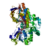
| ||||||||
|---|---|---|---|---|---|---|---|---|---|
| 1 |
| ||||||||
| Unit cell |
|
-Components
| #1: Protein | Mass: 63876.891 Da / Num. of mol.: 1 Source method: isolated from a genetically manipulated source Source: (gene. exp.) Rhodococcus sp. HI-31 (bacteria) / Gene: chnB, chnB1 / Plasmid: pJW234 / Production host: |
|---|---|
| #2: Chemical | ChemComp-FAD /   Mass: 785.550 Da / Num. of mol.: 1 / Source method: obtained synthetically / Formula: C27H33N9O15P2 / Comment: FAD*YM Mass: 785.550 Da / Num. of mol.: 1 / Source method: obtained synthetically / Formula: C27H33N9O15P2 / Comment: FAD*YM |
| #3: Chemical | ChemComp-NAP /   Mass: 743.405 Da / Num. of mol.: 1 / Source method: obtained synthetically / Formula: C21H28N7O17P3 Mass: 743.405 Da / Num. of mol.: 1 / Source method: obtained synthetically / Formula: C21H28N7O17P3 |
| #4: Chemical | ChemComp-CYH /   Mass: 98.143 Da / Num. of mol.: 1 / Source method: obtained synthetically / Formula: C6H10O Mass: 98.143 Da / Num. of mol.: 1 / Source method: obtained synthetically / Formula: C6H10O |
| #5: Water | ChemComp-HOH /  Mass: 18.015 Da / Num. of mol.: 121 / Source method: isolated from a natural source / Formula: H2O Mass: 18.015 Da / Num. of mol.: 121 / Source method: isolated from a natural source / Formula: H2O |
-Experimental details
-Experiment
| Experiment | Method: X-RAY DIFFRACTION / Number of used crystals: 1 |
|---|
- Sample preparation
| Crystal | Density Matthews: 1.92 Å3/Da / Density % sol: 35.83 % |
|---|---|
| Crystal grow | Temperature: 277 K / Method: vapor diffusion, hanging drop / pH: 8 Details: 0.1 M imidazole, 0.2% TMOS, 20% PEG 3350, 0.1 M cyclohexanone, pH 8.0, VAPOR DIFFUSION, HANGING DROP, temperature 277K |
-Data collection
| Diffraction | Mean temperature: 93 K | |||||||||||||||||||||||||||||||||||||||||||||||||||||||||||||||||||||||||||||
|---|---|---|---|---|---|---|---|---|---|---|---|---|---|---|---|---|---|---|---|---|---|---|---|---|---|---|---|---|---|---|---|---|---|---|---|---|---|---|---|---|---|---|---|---|---|---|---|---|---|---|---|---|---|---|---|---|---|---|---|---|---|---|---|---|---|---|---|---|---|---|---|---|---|---|---|---|---|---|
| Diffraction source | Source: ROTATING ANODE / Type: RIGAKU MICROMAX-007 HF / Wavelength: 1.5418 Å | |||||||||||||||||||||||||||||||||||||||||||||||||||||||||||||||||||||||||||||
| Detector | Type: RIGAKU SATURN 944+ / Detector: CCD / Date: Aug 4, 2009 / Details: VariMax HF | |||||||||||||||||||||||||||||||||||||||||||||||||||||||||||||||||||||||||||||
| Radiation | Monochromator: Rotating copper anode / Protocol: SINGLE WAVELENGTH / Monochromatic (M) / Laue (L): M / Scattering type: x-ray | |||||||||||||||||||||||||||||||||||||||||||||||||||||||||||||||||||||||||||||
| Radiation wavelength | Wavelength: 1.5418 Å / Relative weight: 1 | |||||||||||||||||||||||||||||||||||||||||||||||||||||||||||||||||||||||||||||
| Reflection | Resolution: 2.36→50 Å / Num. all: 20262 / Num. obs: 20262 / % possible obs: 97.9 % / Redundancy: 11.2 % / Biso Wilson estimate: 39.4 Å2 / Rmerge(I) obs: 0.087 / Rsym value: 0.087 / Χ2: 1.008 / Net I/σ(I): 9.8 | |||||||||||||||||||||||||||||||||||||||||||||||||||||||||||||||||||||||||||||
| Reflection shell | Diffraction-ID: 1
|
-Phasing
| Phasing | Method: molecular replacement | |||||||||
|---|---|---|---|---|---|---|---|---|---|---|
| Phasing MR | Rfactor: 40.47 / Model details: Phaser MODE: MR_AUTO
|
- Processing
| Software |
| |||||||||||||||||||||||||||||||||||||||||||||||||||||||||||||||||
|---|---|---|---|---|---|---|---|---|---|---|---|---|---|---|---|---|---|---|---|---|---|---|---|---|---|---|---|---|---|---|---|---|---|---|---|---|---|---|---|---|---|---|---|---|---|---|---|---|---|---|---|---|---|---|---|---|---|---|---|---|---|---|---|---|---|---|
| Refinement | Method to determine structure: MOLECULAR REPLACEMENT Starting model: PDB Entry 3GWF Resolution: 2.36→30.61 Å / Cor.coef. Fo:Fc: 0.943 / Cor.coef. Fo:Fc free: 0.903 / WRfactor Rfree: 0.2212 / WRfactor Rwork: 0.1643 / Occupancy max: 1 / Occupancy min: 0.5 / FOM work R set: 0.8073 / SU B: 8.967 / SU ML: 0.213 / SU R Cruickshank DPI: 0.8521 / SU Rfree: 0.3151 / Isotropic thermal model: Isotropic / Cross valid method: THROUGHOUT / σ(F): 0 / ESU R Free: 0.315 / Stereochemistry target values: MAXIMUM LIKELIHOOD / Details: HYDROGENS HAVE BEEN ADDED IN THE RIDING POSITIONS
| |||||||||||||||||||||||||||||||||||||||||||||||||||||||||||||||||
| Solvent computation | Ion probe radii: 0.8 Å / Shrinkage radii: 0.8 Å / VDW probe radii: 1.4 Å / Solvent model: MASK | |||||||||||||||||||||||||||||||||||||||||||||||||||||||||||||||||
| Displacement parameters | Biso max: 65.19 Å2 / Biso mean: 28.4441 Å2 / Biso min: 5.38 Å2
| |||||||||||||||||||||||||||||||||||||||||||||||||||||||||||||||||
| Refinement step | Cycle: LAST / Resolution: 2.36→30.61 Å
| |||||||||||||||||||||||||||||||||||||||||||||||||||||||||||||||||
| Refine LS restraints |
| |||||||||||||||||||||||||||||||||||||||||||||||||||||||||||||||||
| LS refinement shell | Resolution: 2.36→2.425 Å / Total num. of bins used: 20
|
