 Movie
Movie Controller

[English] 日本語
 Yorodumi
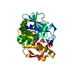
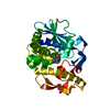
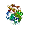
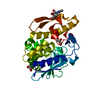
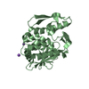
- PDB-3u6z: Crystal structure of the complex formed between type 1 ribosome i... -
+ Open data

- Basic information
| Entry | Database: PDB / ID: 3u6z | ||||||
|---|---|---|---|---|---|---|---|
| Title | Crystal structure of the complex formed between type 1 ribosome inactivating protein and adenine at 1.7A resolution | ||||||
 Components Components | Ribosome inactivating protein | ||||||
 Keywords Keywords | HYDROLASE / RIP / RNA N-glycosidase / Plant protein / nitrogenous base | ||||||
| Function / homology |  Function and homology information Function and homology informationrRNA N-glycosylase / rRNA N-glycosylase activity / defense response / toxin activity / negative regulation of translation / nucleotide binding Similarity search - Function | ||||||
| Biological species |  Momordica balsamina (balsam apple) Momordica balsamina (balsam apple) | ||||||
| Method |  X-RAY DIFFRACTION / SYNCHROTRON / MOLECULAR REPLACEMENT / Resolution: 1.7 Å X-RAY DIFFRACTION / SYNCHROTRON / MOLECULAR REPLACEMENT / Resolution: 1.7 Å | ||||||
 Authors Authors | Pandey, N. / Kushwaha, G.S. / Sinha, M. / Bhushan, A. / Kaur, P. / Sharma, S. / Singh, T.P. | ||||||
 Citation Citation | Journal: Biochim.Biophys.Acta / Year: 2012 Title: Crystal structures of a type-1 ribosome inactivating protein from Momordica balsamina in the bound and unbound states Authors: Kushwaha, G.S. / Pandey, N. / Sinha, M. / Singh, S.B. / Kaur, P. / Sharma, S. / Singh, T.P. | ||||||
| History |
|
- Structure visualization
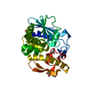
| Structure viewer | Molecule: MolmilJmol/JSmol |
|---|
- Downloads & links
-Download
| PDBx/mmCIF format | 3u6z.cif.gz | 66 KB | Display | PDBx/mmCIF format |
|---|---|---|---|---|
| PDB format | pdb3u6z.ent.gz | 47.5 KB | Display | PDB format |
| PDBx/mmJSON format | 3u6z.json.gz | Tree view | PDBx/mmJSON format | |
| Others |  Other downloads Other downloads |
-Validation report
| Summary document | 3u6z_validation.pdf.gz | 466.4 KB | Display | wwPDB validaton report |
|---|---|---|---|---|
| Full document | 3u6z_full_validation.pdf.gz | 467.1 KB | Display | |
| Data in XML | 3u6z_validation.xml.gz | 13.4 KB | Display | |
| Data in CIF | 3u6z_validation.cif.gz | 19.4 KB | Display | |
| Arichive directory | https://data.pdbj.org/pub/pdb/validation_reports/u6/3u6zftp://data.pdbj.org/pub/pdb/validation_reports/u6/3u6z | HTTPS FTP |
-Related structure data
| Related structure data |  3n1nC  3rl9C 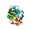 3s9qC  3sj6C 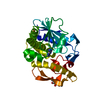 3v2kC 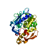 1ahaS S: Starting model for refinement C: citing same article ( |
|---|---|
| Similar structure data |


-Links
 PDBj
PDBj


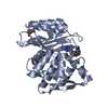
- Assembly
| Deposited unit | 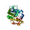
| ||||||||
|---|---|---|---|---|---|---|---|---|---|
| 1 |
| ||||||||
| Unit cell |
|
-Components
| #1: Protein | Mass: 27093.756 Da / Num. of mol.: 1 / Source method: isolated from a natural source / Source: (natural) Momordica balsamina (balsam apple) / References: UniProt: D9J2T9, rRNA N-glycosylase |
|---|---|
| #2: Sugar | ChemComp-NAG /   Type: D-saccharide, beta linking / Mass: 221.208 Da / Num. of mol.: 1 Type: D-saccharide, beta linking / Mass: 221.208 Da / Num. of mol.: 1Source method: isolated from a genetically manipulated source Formula: C8H15NO6 |
| #3: Chemical | ChemComp-ADE /   Mass: 135.127 Da / Num. of mol.: 1 / Source method: obtained synthetically / Formula: C5H5N5 Mass: 135.127 Da / Num. of mol.: 1 / Source method: obtained synthetically / Formula: C5H5N5 |
| #4: Chemical | ChemComp-GOL /   Mass: 92.094 Da / Num. of mol.: 1 / Source method: obtained synthetically / Formula: C3H8O3 Mass: 92.094 Da / Num. of mol.: 1 / Source method: obtained synthetically / Formula: C3H8O3 |
| #5: Water | ChemComp-HOH /  Mass: 18.015 Da / Num. of mol.: 214 / Source method: isolated from a natural source / Formula: H2O Mass: 18.015 Da / Num. of mol.: 214 / Source method: isolated from a natural source / Formula: H2O |
| Has protein modification | Y |
-Experimental details
-Experiment
| Experiment | Method: X-RAY DIFFRACTION / Number of used crystals: 1 |
|---|
- Sample preparation
| Crystal | Density Matthews: 2.4 Å3/Da / Density % sol: 48.71 % |
|---|---|
| Crystal grow | Temperature: 298 K / Method: vapor diffusion, hanging drop / pH: 6.7 Details: 14% PEG 6000, 0.1M Sodium Phosphate, pH 6.7, VAPOR DIFFUSION, HANGING DROP, temperature 298 K |
-Data collection
| Diffraction | Mean temperature: 77 K |
|---|---|
| Diffraction source | Source: SYNCHROTRON / Site: ESRF  / Beamline: BM14 / Wavelength: 0.97 Å / Beamline: BM14 / Wavelength: 0.97 Å |
| Detector | Type: MARRESEARCH / Detector: CCD / Date: Sep 29, 2011 / Details: mirror |
| Radiation | Monochromator: Graphite / Protocol: SINGLE WAVELENGTH / Monochromatic (M) / Laue (L): M / Scattering type: x-ray |
| Radiation wavelength | Wavelength: 0.97 Å / Relative weight: 1 |
| Reflection | Resolution: 1.7→65 Å / Num. all: 26234 / Num. obs: 26234 / % possible obs: 99 % / Observed criterion σ(F): 0 / Observed criterion σ(I): 0 / Biso Wilson estimate: 28.7 Å2 / Rsym value: 0.04 / Net I/σ(I): 47 |
| Reflection shell | Resolution: 1.7→1.74 Å / Mean I/σ(I) obs: 2.2 / Rsym value: 0.618 / % possible all: 99.6 |
- Processing
| Software |
| |||||||||||||||||||||||||||||||||||||||||||||
|---|---|---|---|---|---|---|---|---|---|---|---|---|---|---|---|---|---|---|---|---|---|---|---|---|---|---|---|---|---|---|---|---|---|---|---|---|---|---|---|---|---|---|---|---|---|---|
| Refinement | Method to determine structure: MOLECULAR REPLACEMENT Starting model: PDB ENTRY 1AHA Resolution: 1.7→37.59 Å / Cor.coef. Fo:Fc: 0.973 / Cor.coef. Fo:Fc free: 0.963 / SU B: 2.365 / SU ML: 0.078 / Cross valid method: THROUGHOUT / σ(F): 0 / σ(I): 0 / ESU R Free: 0.104 / Stereochemistry target values: MAXIMUM LIKELIHOOD / Details: HYDROGENS HAVE BEEN USED IF PRESENT IN THE INPUT
| |||||||||||||||||||||||||||||||||||||||||||||
| Solvent computation | Ion probe radii: 0.8 Å / Shrinkage radii: 0.8 Å / VDW probe radii: 1.2 Å / Solvent model: MASK | |||||||||||||||||||||||||||||||||||||||||||||
| Displacement parameters | Biso mean: 33.148 Å2
| |||||||||||||||||||||||||||||||||||||||||||||
| Refinement step | Cycle: LAST / Resolution: 1.7→37.59 Å
| |||||||||||||||||||||||||||||||||||||||||||||
| Refine LS restraints |
| |||||||||||||||||||||||||||||||||||||||||||||
| LS refinement shell | Resolution: 1.7→1.744 Å / Total num. of bins used: 20
|
