- PDB-3tmh: Crystal structure of dual-specific A-kinase anchoring protein 2 i... -
+
Open data
ID or keywords:
Loading...
-
Basic information
Entry
Database: PDB / ID: 3tmh
Title
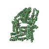
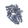
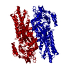
Crystal structure of dual-specific A-kinase anchoring protein 2 in complex with cAMP-dependent protein kinase A type II alpha and PDZK1
Components
A-kinase anchor protein 10, mitochondrial
Na(+)/H(+) exchange regulatory cofactor NHE-RF3
cAMP-dependent protein kinase type II-alpha regulatory subunit
Keywords
TRANSPORT PROTEIN/SIGNALING PROTEIN / alpha helical bundle / PDZ fold / Anchoring protein / PKA / PDZ proteins / membrane / TRANSPORT PROTEIN-SIGNALING PROTEIN complex
Function / homology
Function and homology information
positive regulation of meiotic cell cycle process involved in oocyte maturation / PKA activation in glucagon signalling / CREB1 phosphorylation through the activation of Adenylate Cyclase / DARPP-32 events / carnitine transmembrane transport / GPER1 signaling / Factors involved in megakaryocyte development and platelet production / PKA activation / Hedgehog 'off' state / regulation of meiotic cell cycle process involved in oocyte maturation ...positive regulation of meiotic cell cycle process involved in oocyte maturation / PKA activation in glucagon signalling / CREB1 phosphorylation through the activation of Adenylate Cyclase / DARPP-32 events / carnitine transmembrane transport / GPER1 signaling / Factors involved in megakaryocyte development and platelet production / PKA activation / Hedgehog 'off' state / regulation of meiotic cell cycle process involved in oocyte maturation / scavenger receptor binding / cAMP-dependent protein kinase regulator activity / nucleotide-activated protein kinase complex / xenobiotic detoxification by transmembrane export across the plasma membrane / positive regulation of protein localization to membrane / germinal vesicle / High laminar flow shear stress activates signaling by PIEZO1 and PECAM1:CDH5:KDR in endothelial cells / Vasopressin regulates renal water homeostasis via Aquaporins / cAMP-dependent protein kinase inhibitor activity / beta-2 adrenergic receptor binding / cAMP-dependent protein kinase complex / microvillus membrane / protein kinase A catalytic subunit binding / small molecule binding / protein kinase A binding / brush border / plasma membrane raft / positive regulation of protein targeting to membrane / axoneme / transporter activator activity / cAMP binding / negative regulation of cAMP/PKA signal transduction / protein-membrane adaptor activity / regulation of monoatomic anion transport / T-tubule / protein localization to plasma membrane / PDZ domain binding / brush border membrane / modulation of chemical synaptic transmission / intracellular protein localization / Factors involved in megakaryocyte development and platelet production / adenylate cyclase-activating G protein-coupled receptor signaling pathway / apical plasma membrane / signaling receptor binding / protein domain specific binding / ubiquitin protein ligase binding / centrosome / synapse / protein-containing complex binding / perinuclear region of cytoplasm / glutamatergic synapse / signal transduction / protein-containing complex / mitochondrion / extracellular exosome / identical protein binding / plasma membrane / cytoplasm / cytosol Similarity search - Function
A-kinase anchor protein 10, PKA-binding (AKB) domain / : / : / cAMP-dependent protein kinase regulatory subunit / cAMP-dependent protein kinase regulatory subunit, dimerization-anchoring domain / Regulatory subunit of type II PKA R-subunit / RIIalpha, Regulatory subunit portion of type II PKA R-subunit / : / Regulator of G protein signaling domain / PDZ domain ...A-kinase anchor protein 10, PKA-binding (AKB) domain / : / : / cAMP-dependent protein kinase regulatory subunit / cAMP-dependent protein kinase regulatory subunit, dimerization-anchoring domain / Regulatory subunit of type II PKA R-subunit / RIIalpha, Regulatory subunit portion of type II PKA R-subunit / : / Regulator of G protein signaling domain / PDZ domain / RGS domain / RGS domain profile. / Regulator of G protein signalling domain / RGS, subdomain 2 / RGS domain superfamily / Cyclic nucleotide-binding domain signature 2. / Cyclic nucleotide-binding domain signature 1. / Cyclic nucleotide-binding, conserved site / PDZ domain / Pdz3 Domain / Cyclic nucleotide-monophosphate binding domain / Cyclic nucleotide-binding domain / cAMP/cGMP binding motif profile. / Cyclic nucleotide-binding domain / Cyclic nucleotide-binding domain superfamily / PDZ domain / PDZ domain profile. / Domain present in PSD-95, Dlg, and ZO-1/2. / RmlC-like jelly roll fold / PDZ domain / PDZ superfamily / Roll / Mainly Beta Similarity search - Domain/homology
A-kinase anchor protein 10, mitochondrial / cAMP-dependent protein kinase type II-alpha regulatory subunit / Na(+)/H(+) exchange regulatory cofactor NHE-RF3 Similarity search - Component
Biological species
Homo sapiens (human) Rattus norvegicus (Norway rat)
Mass: 5542.311 Da / Num. of mol.: 4 Fragment: Dimerization/docking domain (D/D), UNP RESIDUES 3-44 Source method: isolated from a genetically manipulated source Source: (gene. exp.) Rattus norvegicus (Norway rat) / Gene: Prkar2a / Production host: Escherichia coli (E. coli) / References: UniProt: P12368
#3: Protein/peptide
A-kinaseanchorprotein10, mitochondrial / AKAP-10 / Dual specificity A kinase-anchoring protein 2 / D-AKAP-2 / Protein kinase A-anchoring ...AKAP-10 / Dual specificity A kinase-anchoring protein 2 / D-AKAP-2 / Protein kinase A-anchoring protein 10 / PRKA10
Mass: 5086.658 Da / Num. of mol.: 3 Fragment: A-kinase binding domain (AKB), UNP RESIDUES 623-662 Source method: isolated from a genetically manipulated source Source: (gene. exp.) Homo sapiens (human) / Gene: AKAP10 / Production host: Escherichia coli (E. coli) / References: UniProt: O43572
In the structure databanks used in Yorodumi, some data are registered as the other names, "COVID-19 virus" and "2019-nCoV". Here are the details of the virus and the list of structure data.
Jan 31, 2019. EMDB accession codes are about to change! (news from PDBe EMDB page)
EMDB accession codes are about to change! (news from PDBe EMDB page)
The allocation of 4 digits for EMDB accession codes will soon come to an end. Whilst these codes will remain in use, new EMDB accession codes will include an additional digit and will expand incrementally as the available range of codes is exhausted. The current 4-digit format prefixed with “EMD-” (i.e. EMD-XXXX) will advance to a 5-digit format (i.e. EMD-XXXXX), and so on. It is currently estimated that the 4-digit codes will be depleted around Spring 2019, at which point the 5-digit format will come into force.
The EM Navigator/Yorodumi systems omit the EMD- prefix.
Related info.:Q: What is EMD? / ID/Accession-code notation in Yorodumi/EM Navigator
Yorodumi is a browser for structure data from EMDB, PDB, SASBDB, etc.
This page is also the successor to EM Navigator detail page, and also detail information page/front-end page for Omokage search.
The word "yorodu" (or yorozu) is an old Japanese word meaning "ten thousand". "mi" (miru) is to see.
Related info.:EMDB / PDB / SASBDB / Comparison of 3 databanks / Yorodumi Search / Aug 31, 2016. New EM Navigator & Yorodumi / Yorodumi Papers / Jmol/JSmol / Function and homology information / Changes in new EM Navigator and Yorodumi
 Movie
Movie Controller

 Yorodumi
 Open data
 Basic information
 Components
 Keywords
 Function and homology information
 Homo sapiens (human)
 X-RAY DIFFRACTION /
  Authors
 Citation
 Structure visualization
 Downloads & links
 Other downloads
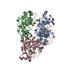


 PDBj
PDBj


 Assembly


 Mass: 18.015 Da / Num. of mol.: 8 / Source method: isolated from a natural source / Formula: H2O
 Sample preparation
 / Beamline: 5.0.1 / Wavelength: 0.9774 Å
 Processing