 Movie
Movie Controller

[English] 日本語
 Yorodumi
- PDB-3tlz: Microcin C7 self immunity protein MccF mutant W186F in complex wi... -
+ Open data

- Basic information
| Entry | Database: PDB / ID: 3tlz | ||||||
|---|---|---|---|---|---|---|---|
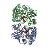
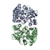
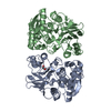
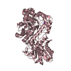
| Title | Microcin C7 self immunity protein MccF mutant W186F in complex with Adenosine Monophosphate | ||||||
 Components Components | MccF | ||||||
 Keywords Keywords | HYDROLASE / serine protease | ||||||
| Function / homology |  Function and homology information Function and homology information | ||||||
| Biological species |  | ||||||
| Method |  X-RAY DIFFRACTION / SYNCHROTRON / MOLECULAR REPLACEMENT / Resolution: 1.5 Å X-RAY DIFFRACTION / SYNCHROTRON / MOLECULAR REPLACEMENT / Resolution: 1.5 Å | ||||||
 Authors Authors | Agarwal, V. / Nair, S.K. | ||||||
 Citation Citation | Journal: Proc.Natl.Acad.Sci.USA / Year: 2012 Title: Structure and function of a serine carboxypeptidase adapted for degradation of the protein synthesis antibiotic microcin C7. Authors: Agarwal, V. / Tikhonov, A. / Metlitskaya, A. / Severinov, K. / Nair, S.K. | ||||||
| History |
|
- Structure visualization
| Structure viewer | Molecule: MolmilJmol/JSmol |
|---|
- Downloads & links
-Download
| PDBx/mmCIF format | 3tlz.cif.gz | 169.2 KB | Display | PDBx/mmCIF format |
|---|---|---|---|---|
| PDB format | pdb3tlz.ent.gz | 128.5 KB | Display | PDB format |
| PDBx/mmJSON format | 3tlz.json.gz | Tree view | PDBx/mmJSON format | |
| Others |  Other downloads Other downloads |
-Validation report
| Arichive directory | https://data.pdbj.org/pub/pdb/validation_reports/tl/3tlzftp://data.pdbj.org/pub/pdb/validation_reports/tl/3tlz | HTTPS FTP |
|---|
-Related structure data
| Related structure data |  3tlaSC 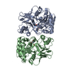 3tlbC  3tlcC  3tleC 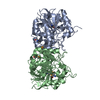 3tlgC  3tlyC S: Starting model for refinement C: citing same article ( |
|---|---|
| Similar structure data |


-Links
 PDBj
PDBj


- Assembly
| Deposited unit | 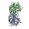
| ||||||||
|---|---|---|---|---|---|---|---|---|---|
| 1 |
| ||||||||
| Unit cell |
|
-Components
| #1: Protein | Mass: 41693.551 Da / Num. of mol.: 2 / Mutation: W186F Source method: isolated from a genetically manipulated source Source: (gene. exp.) #2: Chemical |   Mass: 347.221 Da / Num. of mol.: 2 / Source method: obtained synthetically / Formula: C10H14N5O7P / Comment: AMP*YM Mass: 347.221 Da / Num. of mol.: 2 / Source method: obtained synthetically / Formula: C10H14N5O7P / Comment: AMP*YM#3: Chemical | ChemComp-EDO /   Mass: 62.068 Da / Num. of mol.: 6 / Source method: obtained synthetically / Formula: C2H6O2 Mass: 62.068 Da / Num. of mol.: 6 / Source method: obtained synthetically / Formula: C2H6O2#4: Water | ChemComp-HOH / |  Mass: 18.015 Da / Num. of mol.: 985 / Source method: isolated from a natural source / Formula: H2O Mass: 18.015 Da / Num. of mol.: 985 / Source method: isolated from a natural source / Formula: H2O |
|---|
-Experimental details
-Experiment
| Experiment | Method: X-RAY DIFFRACTION / Number of used crystals: 1 |
|---|
- Sample preparation
| Crystal | Density Matthews: 2.12 Å3/Da / Density % sol: 42 % |
|---|---|
| Crystal grow | Temperature: 282 K / Method: vapor diffusion / pH: 7.5 Details: 0.1M HEPES pH 7.5, 10% PEG 8000, 8% Ethylene Glycol, vapor diffusion, temperature 282K, VAPOR DIFFUSION |
-Data collection
| Diffraction source | Source: SYNCHROTRON / Site: APS  / Beamline: 21-ID-D / Beamline: 21-ID-D |
|---|---|
| Detector | Type: MARMOSAIC 300 mm CCD / Detector: CCD / Date: Jun 5, 2010 |
| Radiation | Protocol: SINGLE WAVELENGTH / Monochromatic (M) / Laue (L): M / Scattering type: x-ray |
| Radiation wavelength | Relative weight: 1 |
| Reflection | Resolution: 1.47→71.8 Å / Num. obs: 115009 / % possible obs: 97.8 % / Redundancy: 5.3 % / Rmerge(I) obs: 0.067 / Rsym value: 0.065 / Net I/σ(I): 38.094 |
| Reflection shell | Resolution: 1.47→1.5 Å / Redundancy: 4.7 % / Rmerge(I) obs: 0.261 / Mean I/σ(I) obs: 5.593 / Rsym value: 0.236 / % possible all: 95 |
- Processing
| Software |
| ||||||||||||||||||||||||||||||||||||||||||||||||||||||||||||||||||||||||||||||||||||||||||||||||||||||||||||||||||||||||||||||||||||||||||||||||||||||||||||||||||||||||||
|---|---|---|---|---|---|---|---|---|---|---|---|---|---|---|---|---|---|---|---|---|---|---|---|---|---|---|---|---|---|---|---|---|---|---|---|---|---|---|---|---|---|---|---|---|---|---|---|---|---|---|---|---|---|---|---|---|---|---|---|---|---|---|---|---|---|---|---|---|---|---|---|---|---|---|---|---|---|---|---|---|---|---|---|---|---|---|---|---|---|---|---|---|---|---|---|---|---|---|---|---|---|---|---|---|---|---|---|---|---|---|---|---|---|---|---|---|---|---|---|---|---|---|---|---|---|---|---|---|---|---|---|---|---|---|---|---|---|---|---|---|---|---|---|---|---|---|---|---|---|---|---|---|---|---|---|---|---|---|---|---|---|---|---|---|---|---|---|---|---|---|---|
| Refinement | Method to determine structure: MOLECULAR REPLACEMENT Starting model: PDB ENTRY 3TLA Resolution: 1.5→25 Å / Cor.coef. Fo:Fc: 0.966 / Cor.coef. Fo:Fc free: 0.958 / Occupancy max: 1 / Occupancy min: 0.5 / SU B: 1.135 / SU ML: 0.044 / Cross valid method: THROUGHOUT / ESU R: 0.076 / ESU R Free: 0.073 / Stereochemistry target values: MAXIMUM LIKELIHOOD / Details: HYDROGENS HAVE BEEN ADDED IN THE RIDING POSITIONS
| ||||||||||||||||||||||||||||||||||||||||||||||||||||||||||||||||||||||||||||||||||||||||||||||||||||||||||||||||||||||||||||||||||||||||||||||||||||||||||||||||||||||||||
| Solvent computation | Ion probe radii: 0.8 Å / Shrinkage radii: 0.8 Å / VDW probe radii: 1.4 Å / Solvent model: BABINET MODEL WITH MASK | ||||||||||||||||||||||||||||||||||||||||||||||||||||||||||||||||||||||||||||||||||||||||||||||||||||||||||||||||||||||||||||||||||||||||||||||||||||||||||||||||||||||||||
| Displacement parameters | Biso mean: 18.818 Å2
| ||||||||||||||||||||||||||||||||||||||||||||||||||||||||||||||||||||||||||||||||||||||||||||||||||||||||||||||||||||||||||||||||||||||||||||||||||||||||||||||||||||||||||
| Refinement step | Cycle: LAST / Resolution: 1.5→25 Å
| ||||||||||||||||||||||||||||||||||||||||||||||||||||||||||||||||||||||||||||||||||||||||||||||||||||||||||||||||||||||||||||||||||||||||||||||||||||||||||||||||||||||||||
| Refine LS restraints |
| ||||||||||||||||||||||||||||||||||||||||||||||||||||||||||||||||||||||||||||||||||||||||||||||||||||||||||||||||||||||||||||||||||||||||||||||||||||||||||||||||||||||||||
| LS refinement shell | Resolution: 1.5→1.539 Å / Total num. of bins used: 20
|
