 Movie
Movie Controller

[English] 日本語
 Yorodumi
- PDB-3sxp: Crystal Structure of Helicobacter pylori ADP-L-glycero-D-manno-he... -
+ Open data

- Basic information
| Entry | Database: PDB / ID: 3sxp | ||||||
|---|---|---|---|---|---|---|---|
| Title | Crystal Structure of Helicobacter pylori ADP-L-glycero-D-manno-heptose-6-epimerase (rfaD, HP0859) | ||||||
 Components Components | ADP-L-glycero-D-mannoheptose-6-epimerase | ||||||
 Keywords Keywords | ISOMERASE / Rossmann fold / Epimerase / NAD binding | ||||||
| Function / homology |  Function and homology information Function and homology informationADP-glyceromanno-heptose 6-epimerase activity / NADP binding / carbohydrate metabolic process Similarity search - Function | ||||||
| Biological species |   Helicobacter pylori (bacteria) Helicobacter pylori (bacteria) | ||||||
| Method |  X-RAY DIFFRACTION / SYNCHROTRON / MOLECULAR REPLACEMENT / Resolution: 2.55 Å X-RAY DIFFRACTION / SYNCHROTRON / MOLECULAR REPLACEMENT / Resolution: 2.55 Å | ||||||
 Authors Authors | Shaik, M.M. / Zanotti, G. / Cendron, L. | ||||||
 Citation Citation | Journal: Biochim.Biophys.Acta / Year: 2011 Title: The crystal structure of ADP-L-glycero-D-manno-heptose-6-epimerase (HP0859) from Helicobacter pylori. Authors: Shaik, M.M. / Zanotti, G. / Cendron, L. #1: Journal: Structure / Year: 2000Title: The crystal structure of ADP-L-glycero-D-mannoheptose 6-epimerase: catalysis with a twist Authors: Deacon, A.M. / Ni, Y.S. / Coleman, W.G. / Ealick, S.E. | ||||||
| History |
|
- Structure visualization
| Structure viewer | Molecule: MolmilJmol/JSmol |
|---|
- Downloads & links
-Download
| PDBx/mmCIF format | 3sxp.cif.gz | 324.4 KB | Display | PDBx/mmCIF format |
|---|---|---|---|---|
| PDB format | pdb3sxp.ent.gz | 263.3 KB | Display | PDB format |
| PDBx/mmJSON format | 3sxp.json.gz | Tree view | PDBx/mmJSON format | |
| Others |  Other downloads Other downloads |
-Validation report
| Summary document | 3sxp_validation.pdf.gz | 1.9 MB | Display | wwPDB validaton report |
|---|---|---|---|---|
| Full document | 3sxp_full_validation.pdf.gz | 2 MB | Display | |
| Data in XML | 3sxp_validation.xml.gz | 67.9 KB | Display | |
| Data in CIF | 3sxp_validation.cif.gz | 89.3 KB | Display | |
| Arichive directory | https://data.pdbj.org/pub/pdb/validation_reports/sx/3sxpftp://data.pdbj.org/pub/pdb/validation_reports/sx/3sxp | HTTPS FTP |
-Related structure data
| Related structure data |  1eq2S S: Starting model for refinement |
|---|---|
| Similar structure data |


-Links
 PDBj
PDBj- Assembly
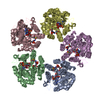
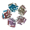
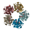
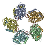
| Deposited unit | 
| ||||||||
|---|---|---|---|---|---|---|---|---|---|
| 1 |
| ||||||||
| Unit cell |
|
-Components
| #1: Protein | Mass: 41147.742 Da / Num. of mol.: 5 / Mutation: G7E Source method: isolated from a genetically manipulated source Source: (gene. exp.) Helicobacter pylori (bacteria) / Strain: G27 / Gene: hp0859, HPG27_813 / Plasmid: pET101-HP0859 / Production host: References: UniProt: B5Z7L9, ADP-glyceromanno-heptose 6-epimerase #2: Chemical | ChemComp-NAD /   Mass: 663.425 Da / Num. of mol.: 5 / Source method: obtained synthetically / Formula: C21H27N7O14P2 / Comment: NAD*YM Mass: 663.425 Da / Num. of mol.: 5 / Source method: obtained synthetically / Formula: C21H27N7O14P2 / Comment: NAD*YM#3: Water | ChemComp-HOH / |  Mass: 18.015 Da / Num. of mol.: 245 / Source method: isolated from a natural source / Formula: H2O Mass: 18.015 Da / Num. of mol.: 245 / Source method: isolated from a natural source / Formula: H2O |
|---|
-Experimental details
-Experiment
| Experiment | Method: X-RAY DIFFRACTION / Number of used crystals: 1 |
|---|
- Sample preparation
| Crystal | Density Matthews: 2.65 Å3/Da / Density % sol: 53.6 % |
|---|---|
| Crystal grow | Temperature: 298 K / Method: vapor diffusion / pH: 5.6 Details: 0.2 M Ammonium Sulphate, 0.1M Tri Sodium citrate pH 5.6, 15% (w/v), PEG 4000, VAPOR DIFFUSION, temperature 298K |
-Data collection
| Diffraction | Mean temperature: 100 K |
|---|---|
| Diffraction source | Source: SYNCHROTRON / Site: ESRF  / Beamline: BM14 / Wavelength: 1 Å / Beamline: BM14 / Wavelength: 1 Å |
| Detector | Type: MAR CCD 165 mm / Detector: CCD / Date: May 5, 2011 |
| Radiation | Protocol: SINGLE WAVELENGTH / Monochromatic (M) / Laue (L): M / Scattering type: x-ray |
| Radiation wavelength | Wavelength: 1 Å / Relative weight: 1 |
| Reflection | Resolution: 2.55→53.38 Å / Num. all: 55485 / Num. obs: 55485 / % possible obs: 86 % / Observed criterion σ(F): 0 / Observed criterion σ(I): 0 / Redundancy: 2.3 % / Biso Wilson estimate: 28.2 Å2 / Rmerge(I) obs: 0.096 / Net I/σ(I): 4.8 |
| Reflection shell | Resolution: 2.55→2.69 Å / Redundancy: 2 % / Rmerge(I) obs: 0.408 / Mean I/σ(I) obs: 1.8 / % possible all: 70.2 |
- Processing
| Software |
| ||||||||||||||||||||||||||||||||||||||||||||||||||||||||||||
|---|---|---|---|---|---|---|---|---|---|---|---|---|---|---|---|---|---|---|---|---|---|---|---|---|---|---|---|---|---|---|---|---|---|---|---|---|---|---|---|---|---|---|---|---|---|---|---|---|---|---|---|---|---|---|---|---|---|---|---|---|---|
| Refinement | Method to determine structure: MOLECULAR REPLACEMENT Starting model: 1EQ2 Resolution: 2.55→53.39 Å / Rfactor Rfree error: 0.005 / Data cutoff high absF: 5954333.06 / Data cutoff low absF: 0 / Isotropic thermal model: RESTRAINED / Cross valid method: THROUGHOUT / σ(F): 0 / σ(I): 0 / Stereochemistry target values: Engh & Huber / Details: BULK SOLVENT MODEL USED
| ||||||||||||||||||||||||||||||||||||||||||||||||||||||||||||
| Solvent computation | Solvent model: FLAT MODEL / Bsol: 72.1885 Å2 / ksol: 0.4 e/Å3 | ||||||||||||||||||||||||||||||||||||||||||||||||||||||||||||
| Displacement parameters | Biso mean: 50.9 Å2
| ||||||||||||||||||||||||||||||||||||||||||||||||||||||||||||
| Refine analyze |
| ||||||||||||||||||||||||||||||||||||||||||||||||||||||||||||
| Refinement step | Cycle: LAST / Resolution: 2.55→53.39 Å
| ||||||||||||||||||||||||||||||||||||||||||||||||||||||||||||
| Refine LS restraints |
| ||||||||||||||||||||||||||||||||||||||||||||||||||||||||||||
| Refine LS restraints NCS | NCS model details: CONSTR | ||||||||||||||||||||||||||||||||||||||||||||||||||||||||||||
| LS refinement shell | Resolution: 2.55→2.71 Å / Rfactor Rfree error: 0.019 / Total num. of bins used: 6
| ||||||||||||||||||||||||||||||||||||||||||||||||||||||||||||
| Xplor file |
|
