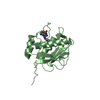
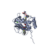
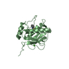
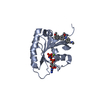
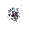
Crystal Structure of Murine Siderocalin (Lipocalin 2, 24p3)
Components
Neutrophil gelatinase-associated lipocalin
Keywords
TRANSPORT PROTEIN / Beta-Barrel / Siderophore binding protein / N-Linked glycosylation / Secreted
Function / homology
Function and homology information
Metal sequestration by antimicrobial proteins / cytoplasmic vesicle lumen / positive regulation of iron ion import across plasma membrane / positive regulation of hippocampal neuron apoptotic process / positive regulation of endothelial tube morphogenesis / negative regulation of hippocampal neuron apoptotic process / positive regulation of cell projection organization / Iron uptake and transport / response to kainic acid / response to mycotoxin ...Metal sequestration by antimicrobial proteins / cytoplasmic vesicle lumen / positive regulation of iron ion import across plasma membrane / positive regulation of hippocampal neuron apoptotic process / positive regulation of endothelial tube morphogenesis / negative regulation of hippocampal neuron apoptotic process / positive regulation of cell projection organization / Iron uptake and transport / response to kainic acid / response to mycotoxin / siderophore transport / cellular response to increased oxygen levels / response to blue light / cellular response to X-ray / response to fructose / short-term memory / cellular response to interleukin-6 / iron ion sequestering activity / enterobactin binding / response to herbicide / response to iron(II) ion / positive regulation of reactive oxygen species biosynthetic process / cellular response to interleukin-1 / long-term memory / extrinsic apoptotic signaling pathway in absence of ligand / Neutrophil degranulation / positive regulation of endothelial cell migration / cellular response to nutrient levels / acute-phase response / response to bacterium / cellular response to nerve growth factor stimulus / cellular response to tumor necrosis factor / positive regulation of reactive oxygen species metabolic process / cellular response to amyloid-beta / response to virus / cellular response to hydrogen peroxide / positive regulation of neuron apoptotic process / positive regulation of cold-induced thermogenesis / cellular response to lipopolysaccharide / protease binding / cellular response to hypoxia / defense response to bacterium / positive regulation of apoptotic process / iron ion binding / response to xenobiotic stimulus / innate immune response / positive regulation of gene expression / : / extracellular region / identical protein binding / cytosol Similarity search - Function
In the structure databanks used in Yorodumi, some data are registered as the other names, "COVID-19 virus" and "2019-nCoV". Here are the details of the virus and the list of structure data.
Jan 31, 2019. EMDB accession codes are about to change! (news from PDBe EMDB page)
EMDB accession codes are about to change! (news from PDBe EMDB page)
The allocation of 4 digits for EMDB accession codes will soon come to an end. Whilst these codes will remain in use, new EMDB accession codes will include an additional digit and will expand incrementally as the available range of codes is exhausted. The current 4-digit format prefixed with “EMD-” (i.e. EMD-XXXX) will advance to a 5-digit format (i.e. EMD-XXXXX), and so on. It is currently estimated that the 4-digit codes will be depleted around Spring 2019, at which point the 5-digit format will come into force.
The EM Navigator/Yorodumi systems omit the EMD- prefix.
Related info.:Q: What is EMD? / ID/Accession-code notation in Yorodumi/EM Navigator
Yorodumi is a browser for structure data from EMDB, PDB, SASBDB, etc.
This page is also the successor to EM Navigator detail page, and also detail information page/front-end page for Omokage search.
The word "yorodu" (or yorozu) is an old Japanese word meaning "ten thousand". "mi" (miru) is to see.
Related info.:EMDB / PDB / SASBDB / Comparison of 3 databanks / Yorodumi Search / Aug 31, 2016. New EM Navigator & Yorodumi / Yorodumi Papers / Jmol/JSmol / Function and homology information / Changes in new EM Navigator and Yorodumi
 Movie
Movie Controller


 Open data
 Basic information
 Components
 Keywords
 Function and homology information
 X-RAY DIFFRACTION /
  Authors
 Citation
 Structure visualization
 Downloads & links
 Other downloads
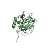


 PDBj
PDBj


 Assembly
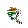
 Homo Sapiens (human) / References: UniProt: P11672
 Mass: 18.015 Da / Num. of mol.: 89 / Source method: isolated from a natural source / Formula: H2O
 Sample preparation
 / Beamline: 5.0.1 / Wavelength: 1 Å
 Processing