[English] 日本語
 Yorodumi
- PDB-3s1a: Crystal structure of the phosphorylation-site double mutant S431E... -
+ Open data
- Basic information
| Entry | Database: PDB / ID: 3s1a | ||||||
|---|---|---|---|---|---|---|---|
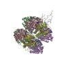
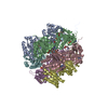
| Title | Crystal structure of the phosphorylation-site double mutant S431E/T432E of the KaiC circadian clock protein | ||||||
 Components Components | (Circadian clock protein kinase kaiC) x 2 | ||||||
 Keywords Keywords | TRANSFERASE / hexamer / atp binding / auto-kinase / phophatase / Serine Threonine Kinase / Mg Binding / Phosphorylation | ||||||
| Function / homology |  Function and homology information Function and homology informationregulation of phosphorelay signal transduction system / negative regulation of circadian rhythm / entrainment of circadian clock / protein serine/threonine/tyrosine kinase activity / circadian rhythm / Hydrolases; Acting on acid anhydrides; Acting on acid anhydrides to facilitate cellular and subcellular movement / non-specific serine/threonine protein kinase / protein serine kinase activity / protein serine/threonine kinase activity / regulation of DNA-templated transcription ...regulation of phosphorelay signal transduction system / negative regulation of circadian rhythm / entrainment of circadian clock / protein serine/threonine/tyrosine kinase activity / circadian rhythm / Hydrolases; Acting on acid anhydrides; Acting on acid anhydrides to facilitate cellular and subcellular movement / non-specific serine/threonine protein kinase / protein serine kinase activity / protein serine/threonine kinase activity / regulation of DNA-templated transcription / magnesium ion binding / ATP hydrolysis activity / DNA binding / ATP binding / identical protein binding Similarity search - Function | ||||||
| Biological species |  Synechococcus elongatus (bacteria) Synechococcus elongatus (bacteria) | ||||||
| Method |  X-RAY DIFFRACTION / X-RAY DIFFRACTION /  SYNCHROTRON / SYNCHROTRON /  MOLECULAR REPLACEMENT / Resolution: 3 Å MOLECULAR REPLACEMENT / Resolution: 3 Å | ||||||
 Authors Authors | Pattanayek, R. / Williams, D.W. / Rossi, G. / Weigand, S. / Mori, T. / Johnson, C.H. / Stewart, P.L. / Egli, M. | ||||||
 Citation Citation |  Journal: Plos One / Year: 2011 Journal: Plos One / Year: 2011Title: Combined SAXS/EM Based Models of the S. elongatus Post-Translational Circadian Oscillator and its Interactions with the Output His-Kinase SasA. Authors: Pattanayek, R. / Williams, D.R. / Rossi, G. / Weigand, S. / Mori, T. / Johnson, C.H. / Stewart, P.L. / Egli, M. | ||||||
| History |
|
- Structure visualization
| Structure viewer | Molecule:  Molmil Molmil Jmol/JSmol Jmol/JSmol |
|---|
- Downloads & links
- Download
| PDBx/mmCIF format |  3s1a.cif.gz 3s1a.cif.gz | 590.5 KB | Display |  PDBx/mmCIF format PDBx/mmCIF format |
|---|---|---|---|---|
| PDB format |  pdb3s1a.ent.gz pdb3s1a.ent.gz | 484.9 KB | Display |  PDB format PDB format |
| PDBx/mmJSON format |  3s1a.json.gz 3s1a.json.gz | Tree view |  PDBx/mmJSON format PDBx/mmJSON format | |
| Others |  Other downloads Other downloads |
-Validation report
| Summary document |  3s1a_validation.pdf.gz 3s1a_validation.pdf.gz | 3.6 MB | Display |  wwPDB validaton report wwPDB validaton report |
|---|---|---|---|---|
| Full document |  3s1a_full_validation.pdf.gz 3s1a_full_validation.pdf.gz | 3.8 MB | Display | |
| Data in XML |  3s1a_validation.xml.gz 3s1a_validation.xml.gz | 135.6 KB | Display | |
| Data in CIF |  3s1a_validation.cif.gz 3s1a_validation.cif.gz | 175.9 KB | Display | |
| Arichive directory |  https://data.pdbj.org/pub/pdb/validation_reports/s1/3s1a https://data.pdbj.org/pub/pdb/validation_reports/s1/3s1a ftp://data.pdbj.org/pub/pdb/validation_reports/s1/3s1a ftp://data.pdbj.org/pub/pdb/validation_reports/s1/3s1a | HTTPS FTP |
-Related structure data
| Related structure data |  3dvlS S: Starting model for refinement |
|---|---|
| Similar structure data |
- Links
Links
- Assembly
| Deposited unit | 
| ||||||||
|---|---|---|---|---|---|---|---|---|---|
| 1 |
| ||||||||
| Unit cell |
|
- Components
| #1: Protein | Mass: 59051.691 Da / Num. of mol.: 2 / Mutation: S431E, T432E Source method: isolated from a genetically manipulated source Details: SEP at residue 320 / Source: (gene. exp.)  Synechococcus elongatus (bacteria) / Strain: PCC 7942 / Gene: kaiC, see0011, Synpcc7942_1216 / Production host: Synechococcus elongatus (bacteria) / Strain: PCC 7942 / Gene: kaiC, see0011, Synpcc7942_1216 / Production host:  References: UniProt: Q79PF4, non-specific serine/threonine protein kinase #2: Protein | Mass: 58971.715 Da / Num. of mol.: 4 / Mutation: S431E, T432E Source method: isolated from a genetically manipulated source Details: SER at residue 320 / Source: (gene. exp.)  Synechococcus elongatus (bacteria) / Strain: PCC 7942 / Gene: kaiC, see0011, Synpcc7942_1216 / Production host: Synechococcus elongatus (bacteria) / Strain: PCC 7942 / Gene: kaiC, see0011, Synpcc7942_1216 / Production host:  References: UniProt: Q79PF4, non-specific serine/threonine protein kinase #3: Chemical | ChemComp-ATP / #4: Chemical | ChemComp-MG / #5: Water | ChemComp-HOH / | Has protein modification | Y | |
|---|
-Experimental details
-Experiment
| Experiment | Method:  X-RAY DIFFRACTION / Number of used crystals: 1 X-RAY DIFFRACTION / Number of used crystals: 1 |
|---|
- Sample preparation
| Crystal | Density Matthews: 2.6 Å3/Da / Density % sol: 52.64 % |
|---|---|
| Crystal grow | Temperature: 291 K / Method: vapor diffusion, hanging drop / pH: 5 Details: SODIUM FORMATE, GLYCEROL, pH 5, VAPOR DIFFUSION, HANGING DROP, temperature 291K |
-Data collection
| Diffraction | Mean temperature: 113.15 K |
|---|---|
| Diffraction source | Source:  SYNCHROTRON / Site: SYNCHROTRON / Site:  APS APS  / Beamline: 21-ID-F / Wavelength: 1 Å / Beamline: 21-ID-F / Wavelength: 1 Å |
| Detector | Type: MARRESEARCH / Detector: CCD / Date: Jan 20, 2009 / Details: mirrors |
| Radiation | Monochromator: SI(111) / Protocol: SINGLE WAVELENGTH / Monochromatic (M) / Laue (L): M / Scattering type: x-ray |
| Radiation wavelength | Wavelength: 1 Å / Relative weight: 1 |
| Reflection | Resolution: 3→17 Å / Num. all: 73982 / Num. obs: 73982 / % possible obs: 99.4 % / Observed criterion σ(F): 0 / Observed criterion σ(I): 0 / Redundancy: 3.9 % / Rmerge(I) obs: 0.075 / Net I/σ(I): 2.3 |
| Reflection shell | Resolution: 3→3.05 Å / Redundancy: 3.9 % / Rmerge(I) obs: 0.381 / Mean I/σ(I) obs: 3.34 / Num. unique all: 3536 / % possible all: 96 |
- Processing
| Software |
| ||||||||||||||||||||
|---|---|---|---|---|---|---|---|---|---|---|---|---|---|---|---|---|---|---|---|---|---|
| Refinement | Method to determine structure:  MOLECULAR REPLACEMENT MOLECULAR REPLACEMENTStarting model: PDB ENTRY 3DVL Resolution: 3→17 Å / σ(F): 2 / Stereochemistry target values: Engh & Huber
| ||||||||||||||||||||
| Refinement step | Cycle: LAST / Resolution: 3→17 Å
| ||||||||||||||||||||
| Refine LS restraints |
| ||||||||||||||||||||
| LS refinement shell | Resolution: 3→3.02 Å
|
 Movie
Movie Controller


 PDBj
PDBj


