 Movie
Movie Controller

[English] 日本語
 Yorodumi
- PDB-3rse: Structural and biochemical characterization of two binding sites ... -
+ Open data

- Basic information
| Entry | Database: PDB / ID: 3rse | ||||||
|---|---|---|---|---|---|---|---|
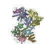
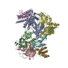
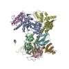
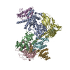
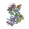
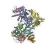
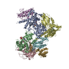
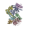
| Title | Structural and biochemical characterization of two binding sites for nucleation promoting factor WASp-VCA on Arp2/3 complex | ||||||
 Components Components |
| ||||||
 Keywords Keywords | STRUCTURAL PROTEIN / Heteroheptamer / Heptameric heterocomplex / F-actin branch initiation / actin / cytosol | ||||||
| Function / homology |  Function and homology information Function and homology informationNOSTRIN mediated eNOS trafficking / Nephrin family interactions / DCC mediated attractive signaling / RHOQ GTPase cycle / muscle cell projection membrane / EPHB-mediated forward signaling / RHOV GTPase cycle / Regulation of actin dynamics for phagocytic cup formation / RHO GTPases Activate WASPs and WAVEs / Arp2/3 protein complex ...NOSTRIN mediated eNOS trafficking / Nephrin family interactions / DCC mediated attractive signaling / RHOQ GTPase cycle / muscle cell projection membrane / EPHB-mediated forward signaling / RHOV GTPase cycle / Regulation of actin dynamics for phagocytic cup formation / RHO GTPases Activate WASPs and WAVEs / Arp2/3 protein complex / Arp2/3 complex-mediated actin nucleation / regulation of actin filament polymerization / Clathrin-mediated endocytosis / dendritic spine morphogenesis / Neutrophil degranulation / cortical cytoskeleton / cilium assembly / positive regulation of double-strand break repair via homologous recombination / positive regulation of lamellipodium assembly / cell projection / actin filament polymerization / structural constituent of cytoskeleton / actin filament binding / cell migration / lamellipodium / synaptic vesicle membrane / site of double-strand break / actin cytoskeleton organization / actin binding / cell cortex / cytoskeleton / protein-macromolecule adaptor activity / postsynapse / endosome / neuron projection / cell division / focal adhesion / endoplasmic reticulum membrane / glutamatergic synapse / positive regulation of transcription by RNA polymerase II / nucleoplasm / ATP binding / identical protein binding / nucleus / cytosol / cytoplasm Similarity search - Function | ||||||
| Biological species |  | ||||||
| Method |  X-RAY DIFFRACTION / SYNCHROTRON / MOLECULAR REPLACEMENT / Resolution: 2.65 Å X-RAY DIFFRACTION / SYNCHROTRON / MOLECULAR REPLACEMENT / Resolution: 2.65 Å | ||||||
 Authors Authors | Pollard, T.D. / Jurgenson, C.T. / Ti, S. / Nolen, B.J. | ||||||
 Citation Citation | Journal: Proc.Natl.Acad.Sci.USA / Year: 2011 Title: Structural and biochemical characterization of two binding sites for nucleation-promoting factor WASp-VCA on Arp2/3 complex. Authors: Ti, S.C. / Jurgenson, C.T. / Nolen, B.J. / Pollard, T.D. | ||||||
| History |
|
- Structure visualization
| Structure viewer | Molecule: MolmilJmol/JSmol |
|---|
- Downloads & links
-Download
| PDBx/mmCIF format | 3rse.cif.gz | 355.1 KB | Display | PDBx/mmCIF format |
|---|---|---|---|---|
| PDB format | pdb3rse.ent.gz | 281.8 KB | Display | PDB format |
| PDBx/mmJSON format | 3rse.json.gz | Tree view | PDBx/mmJSON format | |
| Others |  Other downloads Other downloads |
-Validation report
| Arichive directory | https://data.pdbj.org/pub/pdb/validation_reports/rs/3rseftp://data.pdbj.org/pub/pdb/validation_reports/rs/3rse | HTTPS FTP |
|---|
-Related structure data
| Related structure data |  2p9sS S: Starting model for refinement |
|---|---|
| Similar structure data |


-Links
 PDBj
PDBj


- Assembly
| Deposited unit | 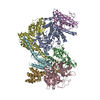
| ||||||||
|---|---|---|---|---|---|---|---|---|---|
| 1 |
| ||||||||
| Unit cell |
|
-Components
-Actin-related protein ... , 7 types, 7 molecules ABCDEFG
| #1: Protein | Mass: 47428.031 Da / Num. of mol.: 1 / Source method: isolated from a natural source / Source: (natural) |
|---|---|
| #2: Protein | Mass: 44818.711 Da / Num. of mol.: 1 / Source method: isolated from a natural source / Source: (natural) |
| #3: Protein | Mass: 41030.766 Da / Num. of mol.: 1 / Source method: isolated from a natural source / Source: (natural) |
| #4: Protein | Mass: 34402.043 Da / Num. of mol.: 1 / Source method: isolated from a natural source / Source: (natural) |
| #5: Protein | Mass: 20572.666 Da / Num. of mol.: 1 / Source method: isolated from a natural source / Source: (natural) |
| #6: Protein | Mass: 19697.047 Da / Num. of mol.: 1 / Source method: isolated from a natural source / Source: (natural) |
| #7: Protein | Mass: 16251.308 Da / Num. of mol.: 1 / Source method: isolated from a natural source / Source: (natural) |
-Protein/peptide / Non-polymers , 2 types, 209 molecules Z
| #8: Protein/peptide | Mass: 462.453 Da / Num. of mol.: 1 Source method: isolated from a genetically manipulated source Source: (gene. exp.)  |
|---|---|
| #9: Water | ChemComp-HOH / Mass: 18.015 Da / Num. of mol.: 208 / Source method: isolated from a natural source / Formula: H2O |
-Experimental details
-Experiment
| Experiment | Method: X-RAY DIFFRACTION / Number of used crystals: 1 |
|---|
- Sample preparation
| Crystal | Density Matthews: 3.28 Å3/Da / Density % sol: 62.51 % |
|---|---|
| Crystal grow | Temperature: 277 K / Method: vapor diffusion, hanging drop / pH: 7.5 Details: 100 mM HEPES 7.5, 8% PEG 8000, 200 mM KCl, VAPOR DIFFUSION, HANGING DROP, temperature 277K |
-Data collection
| Diffraction | Mean temperature: 100 K |
|---|---|
| Diffraction source | Source: SYNCHROTRON / Site: NSLS  / Beamline: X25 / Wavelength: 1 Å / Beamline: X25 / Wavelength: 1 Å |
| Detector | Type: ADSC QUANTUM 315 / Detector: CCD / Date: Aug 15, 2009 |
| Radiation | Protocol: SINGLE WAVELENGTH / Monochromatic (M) / Laue (L): M / Scattering type: x-ray |
| Radiation wavelength | Wavelength: 1 Å / Relative weight: 1 |
| Reflection | Resolution: 2.65→50 Å / Num. all: 87036 / Num. obs: 87036 / Redundancy: 5.3 % / Rmerge(I) obs: 0.09 / Rsym value: 0.09 / Net I/σ(I): 23.9 |
| Reflection shell | Resolution: 2.65→2.74 Å / Redundancy: 5.1 % / Rmerge(I) obs: 0.817 / Mean I/σ(I) obs: 1 / Num. unique all: 8474 |
- Processing
| Software |
| ||||||||||||||||||||
|---|---|---|---|---|---|---|---|---|---|---|---|---|---|---|---|---|---|---|---|---|---|
| Refinement | Method to determine structure: MOLECULAR REPLACEMENT Starting model: PDB entry 2P9S Resolution: 2.65→50 Å / Cross valid method: THROUGHOUT / σ(F): 1 / Stereochemistry target values: Engh & Huber
| ||||||||||||||||||||
| Refinement step | Cycle: LAST / Resolution: 2.65→50 Å
| ||||||||||||||||||||
| LS refinement shell | Resolution: 2.65→2.71 Å
|
