 Movie
Movie Controller

[English] 日本語
 Yorodumi
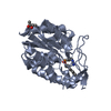
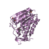
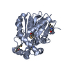
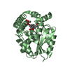
- PDB-3rli: Crystal structure of monoacylglycerol lipase from Bacillus sp. H2... -
+ Open data

- Basic information
| Entry | Database: PDB / ID: 3rli | ||||||
|---|---|---|---|---|---|---|---|
| Title | Crystal structure of monoacylglycerol lipase from Bacillus sp. H257 in complex with PMSF | ||||||
 Components Components | Thermostable monoacylglycerol lipase | ||||||
 Keywords Keywords | HYDROLASE / alpha/beta hydrolase | ||||||
| Function / homology |  Function and homology information Function and homology information | ||||||
| Biological species |  | ||||||
| Method |  X-RAY DIFFRACTION / SYNCHROTRON / MOLECULAR REPLACEMENT / molecular replacement / Resolution: 1.854 Å X-RAY DIFFRACTION / SYNCHROTRON / MOLECULAR REPLACEMENT / molecular replacement / Resolution: 1.854 Å | ||||||
 Authors Authors | Rengachari, S. / Bezerra, G.A. / Gruber, K. / Oberer, M. | ||||||
 Citation Citation | Journal: Biochim.Biophys.Acta / Year: 2012 Title: The structure of monoacylglycerol lipase from Bacillus sp. H257 reveals unexpected conservation of the cap architecture between bacterial and human enzymes. Authors: Rengachari, S. / Bezerra, G.A. / Riegler-Berket, L. / Gruber, C.C. / Sturm, C. / Taschler, U. / Boeszoermenyi, A. / Dreveny, I. / Zimmermann, R. / Gruber, K. / Oberer, M. | ||||||
| History |
|
- Structure visualization
| Structure viewer | Molecule: MolmilJmol/JSmol |
|---|
- Downloads & links
-Download
| PDBx/mmCIF format | 3rli.cif.gz | 67 KB | Display | PDBx/mmCIF format |
|---|---|---|---|---|
| PDB format | pdb3rli.ent.gz | 47.1 KB | Display | PDB format |
| PDBx/mmJSON format | 3rli.json.gz | Tree view | PDBx/mmJSON format | |
| Others |  Other downloads Other downloads |
-Validation report
| Arichive directory | https://data.pdbj.org/pub/pdb/validation_reports/rl/3rliftp://data.pdbj.org/pub/pdb/validation_reports/rl/3rli | HTTPS FTP |
|---|
-Related structure data
| Related structure data | 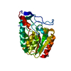 3rm3SC S: Starting model for refinement C: citing same article ( |
|---|---|
| Similar structure data |
-Links
 PDBj
PDBj- Assembly
| Deposited unit | 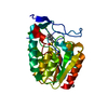
| ||||||||
|---|---|---|---|---|---|---|---|---|---|
| 1 |
| ||||||||
| Unit cell |
|
-Components
| #1: Protein | Mass: 29560.611 Da / Num. of mol.: 1 Source method: isolated from a genetically manipulated source Source: (gene. exp.) |
|---|---|
| #2: Chemical | ChemComp-MRD / (  Mass: 118.174 Da / Num. of mol.: 1 / Source method: obtained synthetically / Formula: C6H14O2 / Comment: precipitant*YM Mass: 118.174 Da / Num. of mol.: 1 / Source method: obtained synthetically / Formula: C6H14O2 / Comment: precipitant*YM |
| #3: Chemical | ChemComp-PMS /   Mass: 172.202 Da / Num. of mol.: 1 / Source method: obtained synthetically / Formula: C7H8O3S Mass: 172.202 Da / Num. of mol.: 1 / Source method: obtained synthetically / Formula: C7H8O3S |
| #4: Water | ChemComp-HOH /  Mass: 18.015 Da / Num. of mol.: 202 / Source method: isolated from a natural source / Formula: H2O Mass: 18.015 Da / Num. of mol.: 202 / Source method: isolated from a natural source / Formula: H2O |
| Has protein modification | Y |
-Experimental details
-Experiment
| Experiment | Method: X-RAY DIFFRACTION / Number of used crystals: 1 |
|---|
- Sample preparation
| Crystal | Density Matthews: 1.84 Å3/Da / Density % sol: 33.04 % |
|---|---|
| Crystal grow | Temperature: 293 K / Method: vapor diffusion, sitting drop / pH: 6.5 Details: 0.1 M MES/imidazole pH 6.5, 12.5% w/v PEG 1000, 12.5% w/v PEG 3350, 12.5% v/v MPD, and 0.02 M of monosaccharides (D-glucose, D-mannose, D-galactose, L-fuctose, D-xylose, and N-acetyl-D- ...Details: 0.1 M MES/imidazole pH 6.5, 12.5% w/v PEG 1000, 12.5% w/v PEG 3350, 12.5% v/v MPD, and 0.02 M of monosaccharides (D-glucose, D-mannose, D-galactose, L-fuctose, D-xylose, and N-acetyl-D-glucosamine), VAPOR DIFFUSION, SITTING DROP, temperature 293.0K |
-Data collection
| Diffraction | Mean temperature: 100 K |
|---|---|
| Diffraction source | Source: SYNCHROTRON / Site: EMBL/DESY, HAMBURG  / Beamline: X13 / Wavelength: 0.81 Å / Beamline: X13 / Wavelength: 0.81 Å |
| Detector | Type: MAR CCD 165 mm / Detector: CCD / Date: Oct 31, 2010 |
| Radiation | Monochromator: Si 111, horizontally focussing / Protocol: SINGLE WAVELENGTH / Monochromatic (M) / Laue (L): M / Scattering type: x-ray |
| Radiation wavelength | Wavelength: 0.81 Å / Relative weight: 1 |
| Reflection | Resolution: 1.854→40.376 Å / Num. all: 17878 / Num. obs: 17878 / % possible obs: 96.2 % / Redundancy: 3.1 % / Biso Wilson estimate: 15.56 Å2 / Rsym value: 0.057 |
| Reflection shell | Resolution: 1.854→1.94 Å / % possible all: 96.2 |
-Phasing
| Phasing | Method: molecular replacement | |||||||||
|---|---|---|---|---|---|---|---|---|---|---|
| Phasing MR | Rfactor: 27.63
|
- Processing
| Software |
| |||||||||||||||||||||||||||||||||||||||||||||||||
|---|---|---|---|---|---|---|---|---|---|---|---|---|---|---|---|---|---|---|---|---|---|---|---|---|---|---|---|---|---|---|---|---|---|---|---|---|---|---|---|---|---|---|---|---|---|---|---|---|---|---|
| Refinement | Method to determine structure: MOLECULAR REPLACEMENT Starting model: 3RM3 Resolution: 1.854→17.683 Å / Occupancy max: 1 / Occupancy min: 0.46 / FOM work R set: 0.8828 / SU ML: 0.21 / σ(F): 0 / Phase error: 18.98 / Stereochemistry target values: ML
| |||||||||||||||||||||||||||||||||||||||||||||||||
| Solvent computation | Shrinkage radii: 0.83 Å / VDW probe radii: 1.1 Å / Solvent model: FLAT BULK SOLVENT MODEL / Bsol: 59.738 Å2 / ksol: 0.437 e/Å3 | |||||||||||||||||||||||||||||||||||||||||||||||||
| Displacement parameters | Biso max: 71.65 Å2 / Biso mean: 17.4197 Å2 / Biso min: 4.06 Å2
| |||||||||||||||||||||||||||||||||||||||||||||||||
| Refinement step | Cycle: LAST / Resolution: 1.854→17.683 Å
| |||||||||||||||||||||||||||||||||||||||||||||||||
| Refine LS restraints |
| |||||||||||||||||||||||||||||||||||||||||||||||||
| LS refinement shell | Refine-ID: X-RAY DIFFRACTION / Total num. of bins used: 6
|
