 Movie
Movie Controller

[English] 日本語
 Yorodumi
- PDB-3rlc: Crystal structure of the read-through domain from bacteriophage Q... -
+ Open data

- Basic information
| Entry | Database: PDB / ID: 3rlc | ||||||
|---|---|---|---|---|---|---|---|
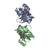
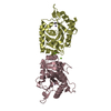
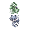
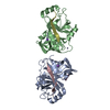
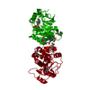
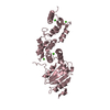
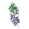
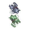
| Title | Crystal structure of the read-through domain from bacteriophage Qbeta A1 protein, hexagonal crystal form | ||||||
 Components Components | A1 protein | ||||||
 Keywords Keywords | STRUCTURAL PROTEIN / Beta-barrel / polyproline helix | ||||||
| Function / homology | Read-through domain / Read-through domain / Levivirus coat protein / Levivirus coat protein / Bacteriophage RNA-type, capsid / viral capsid / structural molecule activity / Minor capsid protein A1 Function and homology information Function and homology information | ||||||
| Biological species |  Enterobacteria phage Qbeta (virus) Enterobacteria phage Qbeta (virus) | ||||||
| Method |  X-RAY DIFFRACTION / SYNCHROTRON / MOLECULAR REPLACEMENT / molecular replacement / Resolution: 2.9 Å X-RAY DIFFRACTION / SYNCHROTRON / MOLECULAR REPLACEMENT / molecular replacement / Resolution: 2.9 Å | ||||||
 Authors Authors | Rumnieks, J. / Tars, K. | ||||||
 Citation Citation | Journal: Protein Sci. / Year: 2011 Title: Crystal structure of the read-through domain from bacteriophage Qbeta A1 protein Authors: Rumnieks, J. / Tars, K. | ||||||
| History |
|
- Structure visualization
| Structure viewer | Molecule: MolmilJmol/JSmol |
|---|
- Downloads & links
-Download
| PDBx/mmCIF format | 3rlc.cif.gz | 49.9 KB | Display | PDBx/mmCIF format |
|---|---|---|---|---|
| PDB format | pdb3rlc.ent.gz | 34.7 KB | Display | PDB format |
| PDBx/mmJSON format | 3rlc.json.gz | Tree view | PDBx/mmJSON format | |
| Others |  Other downloads Other downloads |
-Validation report
| Arichive directory | https://data.pdbj.org/pub/pdb/validation_reports/rl/3rlcftp://data.pdbj.org/pub/pdb/validation_reports/rl/3rlc | HTTPS FTP |
|---|
-Related structure data


-Links
 PDBj
PDBj- Assembly
| Deposited unit | 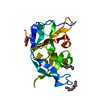
| ||||||||
|---|---|---|---|---|---|---|---|---|---|
| 1 | 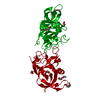
| ||||||||
| 2 |
| ||||||||
| Unit cell |
|
-Components
| #1: Protein | Mass: 21794.596 Da / Num. of mol.: 1 / Fragment: Read-through domain, UNP residues 145-329 Source method: isolated from a genetically manipulated source Source: (gene. exp.) Enterobacteria phage Qbeta (virus) / Gene: A1 / Plasmid: pBAD / Production host:  | ||
|---|---|---|---|
| #2: Chemical | ChemComp-PG4 /   Mass: 194.226 Da / Num. of mol.: 4 / Source method: obtained synthetically / Formula: C8H18O5 / Comment: precipitant*YM Mass: 194.226 Da / Num. of mol.: 4 / Source method: obtained synthetically / Formula: C8H18O5 / Comment: precipitant*YM#3: Water | ChemComp-HOH / |  Mass: 18.015 Da / Num. of mol.: 20 / Source method: isolated from a natural source / Formula: H2O Mass: 18.015 Da / Num. of mol.: 20 / Source method: isolated from a natural source / Formula: H2O |
-Experimental details
-Experiment
| Experiment | Method: X-RAY DIFFRACTION / Number of used crystals: 1 |
|---|
- Sample preparation
| Crystal | Density Matthews: 2.65 Å3/Da / Density % sol: 53.51 % |
|---|---|
| Crystal grow | Temperature: 298 K / Method: vapor diffusion, sitting drop / pH: 8 Details: 40% PEG 300, pH 8.0, VAPOR DIFFUSION, SITTING DROP, temperature 298K |
-Data collection
| Diffraction | Mean temperature: 100 K | ||||||||||||||||||||||||||||||||||||||||||||||||||||||||||||||||||||||||||||||||||||||||
|---|---|---|---|---|---|---|---|---|---|---|---|---|---|---|---|---|---|---|---|---|---|---|---|---|---|---|---|---|---|---|---|---|---|---|---|---|---|---|---|---|---|---|---|---|---|---|---|---|---|---|---|---|---|---|---|---|---|---|---|---|---|---|---|---|---|---|---|---|---|---|---|---|---|---|---|---|---|---|---|---|---|---|---|---|---|---|---|---|---|
| Diffraction source | Source: SYNCHROTRON / Site: MAX II  / Beamline: I911-2 / Wavelength: 1.03874 Å / Beamline: I911-2 / Wavelength: 1.03874 Å | ||||||||||||||||||||||||||||||||||||||||||||||||||||||||||||||||||||||||||||||||||||||||
| Detector | Type: MAR CCD 165 mm / Detector: CCD / Date: Feb 17, 2011 | ||||||||||||||||||||||||||||||||||||||||||||||||||||||||||||||||||||||||||||||||||||||||
| Radiation | Monochromator: Bent Si (111) crystal, horizontally focusing / Protocol: SINGLE WAVELENGTH / Monochromatic (M) / Laue (L): M / Scattering type: x-ray | ||||||||||||||||||||||||||||||||||||||||||||||||||||||||||||||||||||||||||||||||||||||||
| Radiation wavelength | Wavelength: 1.03874 Å / Relative weight: 1 | ||||||||||||||||||||||||||||||||||||||||||||||||||||||||||||||||||||||||||||||||||||||||
| Reflection | Resolution: 2.9→34.555 Å / Num. obs: 5773 / % possible obs: 100 % / Observed criterion σ(F): 1.4 / Observed criterion σ(I): 1.4 / Redundancy: 9.5 % / Rsym value: 0.098 / Net I/σ(I): 17.5 | ||||||||||||||||||||||||||||||||||||||||||||||||||||||||||||||||||||||||||||||||||||||||
| Reflection shell | Diffraction-ID: 1
|
-Phasing
| Phasing | Method: molecular replacement | |||||||||
|---|---|---|---|---|---|---|---|---|---|---|
| Phasing MR |
|
- Processing
| Software |
| |||||||||||||||||||||||||||||||||||||||||||||||||||||||||||||||||
|---|---|---|---|---|---|---|---|---|---|---|---|---|---|---|---|---|---|---|---|---|---|---|---|---|---|---|---|---|---|---|---|---|---|---|---|---|---|---|---|---|---|---|---|---|---|---|---|---|---|---|---|---|---|---|---|---|---|---|---|---|---|---|---|---|---|---|
| Refinement | Method to determine structure: MOLECULAR REPLACEMENT / Resolution: 2.9→29.93 Å / Cor.coef. Fo:Fc: 0.926 / Cor.coef. Fo:Fc free: 0.884 / WRfactor Rfree: 0.2542 / WRfactor Rwork: 0.1817 / Occupancy max: 1 / Occupancy min: 1 / FOM work R set: 0.7806 / SU B: 18.817 / SU ML: 0.355 / SU Rfree: 0.4537 / Cross valid method: THROUGHOUT / σ(F): 0 / ESU R Free: 0.454 / Stereochemistry target values: MAXIMUM LIKELIHOOD Details: HYDROGENS HAVE BEEN ADDED IN THE RIDING POSITIONS U VALUES: REFINED INDIVIDUALLY
| |||||||||||||||||||||||||||||||||||||||||||||||||||||||||||||||||
| Solvent computation | Ion probe radii: 0.8 Å / Shrinkage radii: 0.8 Å / VDW probe radii: 1.4 Å / Solvent model: MASK | |||||||||||||||||||||||||||||||||||||||||||||||||||||||||||||||||
| Displacement parameters | Biso max: 67.75 Å2 / Biso mean: 41.5926 Å2 / Biso min: 16.93 Å2
| |||||||||||||||||||||||||||||||||||||||||||||||||||||||||||||||||
| Refinement step | Cycle: LAST / Resolution: 2.9→29.93 Å
| |||||||||||||||||||||||||||||||||||||||||||||||||||||||||||||||||
| Refine LS restraints |
| |||||||||||||||||||||||||||||||||||||||||||||||||||||||||||||||||
| LS refinement shell | Resolution: 2.9→2.976 Å / Total num. of bins used: 20
|
