- PDB-3rb8: Structure of the phage tubulin PhuZ(SeMet)-GDP -
+
Open data
ID or keywords:
Loading...
-
Basic information
Entry
Database: PDB / ID: 3rb8
Title
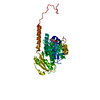
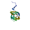
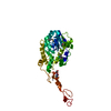
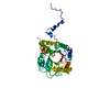
Structure of the phage tubulin PhuZ(SeMet)-GDP
Components
Putative uncharacterized protein
Keywords
UNKNOWN FUNCTION / Tubulin
Function / homology
Function and homology information
positive regulation of viral life cycle / host cell cytoplasm / Hydrolases; Acting on acid anhydrides; Acting on GTP to facilitate cellular and subcellular movement / GTPase activity / GTP binding / identical protein binding Similarity search - Function
Method to determine structure: MAD / Resolution: 2.6→46.379 Å / SU ML: 0.34 Isotropic thermal model: Isotropic with TLS refinement with eleven groups σ(F): 1 / Stereochemistry target values: ML
Rfactor
Num. reflection
% reflection
Selection details
Rfree
0.2373
1867
9.92 %
random
Rwork
0.1633
-
-
-
obs
0.1706
18830
95.32 %
-
all
-
23023
-
-
Solvent computation
Shrinkage radii: 0.9 Å / VDW probe radii: 1.11 Å / Solvent model: FLAT BULK SOLVENT MODEL / Bsol: 40.427 Å2 / ksol: 0.313 e/Å3
Displacement parameters
Baniso -1
Baniso -2
Baniso -3
1-
-5.2866 Å2
-0 Å2
0 Å2
2-
-
-3.2448 Å2
-0 Å2
3-
-
-
8.5313 Å2
Refinement step
Cycle: LAST / Resolution: 2.6→46.379 Å
Protein
Nucleic acid
Ligand
Solvent
Total
Num. atoms
2419
0
29
14
2462
Refine LS restraints
Refine-ID
Type
Dev ideal
Number
X-RAY DIFFRACTION
f_bond_d
0.008
2487
X-RAY DIFFRACTION
f_angle_d
1.141
3385
X-RAY DIFFRACTION
f_dihedral_angle_d
14.645
922
X-RAY DIFFRACTION
f_chiral_restr
0.07
404
X-RAY DIFFRACTION
f_plane_restr
0.004
435
LS refinement shell
Refine-ID: X-RAY DIFFRACTION
Resolution (Å)
Rfactor Rfree
Num. reflection Rfree
Rfactor Rwork
Num. reflection Rwork
Num. reflection obs
% reflection obs (%)
2.6-2.6705
0.3415
123
0.2491
1181
1181
85
2.6705-2.749
0.3643
134
0.2537
1203
1203
88
2.749-2.8377
0.3505
133
0.2336
1223
1223
90
2.8377-2.9392
0.3427
138
0.2158
1253
1253
91
2.9392-3.0568
0.2815
136
0.2125
1271
1271
94
3.0568-3.1959
0.3134
147
0.2061
1332
1332
97
3.1959-3.3643
0.324
151
0.1935
1360
1360
99
3.3643-3.5751
0.249
154
0.1838
1346
1346
98
3.5751-3.851
0.2044
157
0.15
1329
1329
99
3.851-4.2383
0.2244
141
0.1345
1376
1376
99
4.2383-4.851
0.1678
150
0.1095
1348
1348
100
4.851-6.1095
0.1969
147
0.1451
1378
1378
100
6.1095-46.3864
0.2029
156
0.1487
1363
1363
100
Refinement TLS params.
Method: refined / Refine-ID: X-RAY DIFFRACTION
ID
L11 (°2)
L12 (°2)
L13 (°2)
L22 (°2)
L23 (°2)
L33 (°2)
S11 (Å °)
S12 (Å °)
S13 (Å °)
S21 (Å °)
S22 (Å °)
S23 (Å °)
S31 (Å °)
S32 (Å °)
S33 (Å °)
T11 (Å2)
T12 (Å2)
T13 (Å2)
T22 (Å2)
T23 (Å2)
T33 (Å2)
Origin x (Å)
Origin y (Å)
Origin z (Å)
1
0.4376
-0.1173
0.1835
0.2989
0.0534
0.3723
-0.2409
-0.0226
0.0966
0.375
0.0152
-0.2567
-0.4987
0.1391
-0.0396
0.6371
0.0575
-0.3866
0.2404
-0.0387
0.6271
4.4315
4.7768
-1.7166
2
0.2574
0.1223
-0.4037
0.9652
-0.9981
1.3724
-0.6739
0.0922
1.0339
-0.1421
-0.2885
-0.2168
-0.8433
0.1541
-0.3363
1.0642
0.1336
-0.6223
0.6127
-0.1859
1.0126
15.3378
1.2578
2.774
3
0.3878
-0.1716
0.1849
1.2592
-0.6157
0.3549
-0.1435
-0.3146
0.1726
0.7655
0.3376
-0.1374
-0.3441
-0.083
0.0542
0.6524
0.1498
-0.1345
0.4547
0.0115
0.337
5.8419
-7.5469
0.4418
4
1.1826
-0.1424
0.3951
1.3256
0.3443
0.3878
-0.0761
0.1502
0.052
0.4865
0.1355
-0.011
0.3279
0.097
-0.0291
0.3654
0.0489
-0.0838
0.2277
0.0166
0.2402
5.6162
-10.2373
-9.1494
5
0.0576
0.1788
0.0959
0.9841
-0.3975
1.3251
-0.2244
-0.142
0.0535
-0.0748
-0.0012
-0.3685
0.0384
0.1337
0.1099
0.4147
-0.0609
0.0416
0.4572
0.0634
0.3691
4.4398
-4.8005
-16.0528
6
0.0472
-0.2227
0.0754
1.0535
-0.3535
0.3613
-0.1019
-0.0518
0.2332
0.0428
-0.1802
-0.7389
-0.1497
0.2202
-0.3389
0.8194
-0.0155
-0.4026
-0.0133
0.1721
0.6451
3.3084
7.4044
-13.46
7
0.3312
-0.1326
0.3471
1.5603
0.4785
0.633
0.0613
-0.4202
-0.1868
-0.1545
-0.1649
1.0436
0.0081
-0.4157
0.063
0.3522
-0.0259
0.0556
0.6025
0.0397
0.6554
-12.403
-4.6901
-12.2029
8
0.5
-0.0394
-0.4166
0.9815
-0.1673
0.4134
-0.7129
0.4377
0.3982
-0.7006
-0.2493
0.088
-0.7716
0.1361
-1.1284
0.5746
0.0381
-0.6264
0.3561
0.1298
0.6366
-12.1523
3.536
-23.9418
9
1.3228
-0.51
-0.5542
1.2904
-0.341
0.5104
-0.4024
-0.0795
0.4734
-0.3468
0.0751
0.5228
-0.575
-0.5213
-0.5398
0.4966
0.1734
-0.2825
0.4976
-0.0677
0.6915
-13.2321
5.7334
-18.2082
10
0.4177
0.0838
-0.4826
0.9921
0.3668
1.5117
-0.3794
0.4884
0.1639
-0.1978
0.2387
-0.5352
-0.0032
0.2427
0.0568
0.4508
-0.1126
0.0226
0.6621
-0.0905
0.4001
15.431
-13.4639
-24.709
11
2.3849
-1.1648
-0.1854
2.991
0.1304
0.5958
-0.1396
0.3501
-0.5466
0.9078
0.1846
0.5487
0.439
-0.1196
0.0286
0.5422
-0.0496
-0.0752
0.5183
0.0672
0.5026
49.9479
-20.4309
-13.0733
Refinement TLS group
ID
Refine-ID
Refine TLS-ID
Selection details
1
X-RAY DIFFRACTION
1
CHAINAAND (RESSEQ2:32)
2
X-RAY DIFFRACTION
2
CHAINAAND (RESSEQ33:59)
3
X-RAY DIFFRACTION
3
CHAINAAND (RESSEQ60:94)
4
X-RAY DIFFRACTION
4
CHAINAAND (RESSEQ95:143)
5
X-RAY DIFFRACTION
5
CHAINAAND (RESSEQ144:160)
6
X-RAY DIFFRACTION
6
CHAINAAND (RESSEQ161:178)
7
X-RAY DIFFRACTION
7
CHAINAAND (RESSEQ179:214)
8
X-RAY DIFFRACTION
8
CHAINAAND (RESSEQ215:238)
9
X-RAY DIFFRACTION
9
CHAINAAND (RESSEQ239:271)
10
X-RAY DIFFRACTION
10
CHAINAAND (RESSEQ272:294)
11
X-RAY DIFFRACTION
11
CHAINAAND (RESSEQ295:315)
+
About Yorodumi
-
News
-
Feb 9, 2022. New format data for meta-information of EMDB entries
New format data for meta-information of EMDB entries
Version 3 of the EMDB header file is now the official format.
The previous official version 1.9 will be removed from the archive.
In the structure databanks used in Yorodumi, some data are registered as the other names, "COVID-19 virus" and "2019-nCoV". Here are the details of the virus and the list of structure data.
Jan 31, 2019. EMDB accession codes are about to change! (news from PDBe EMDB page)
EMDB accession codes are about to change! (news from PDBe EMDB page)
The allocation of 4 digits for EMDB accession codes will soon come to an end. Whilst these codes will remain in use, new EMDB accession codes will include an additional digit and will expand incrementally as the available range of codes is exhausted. The current 4-digit format prefixed with “EMD-” (i.e. EMD-XXXX) will advance to a 5-digit format (i.e. EMD-XXXXX), and so on. It is currently estimated that the 4-digit codes will be depleted around Spring 2019, at which point the 5-digit format will come into force.
The EM Navigator/Yorodumi systems omit the EMD- prefix.
Related info.:Q: What is EMD? / ID/Accession-code notation in Yorodumi/EM Navigator
Yorodumi is a browser for structure data from EMDB, PDB, SASBDB, etc.
This page is also the successor to EM Navigator detail page, and also detail information page/front-end page for Omokage search.
The word "yorodu" (or yorozu) is an old Japanese word meaning "ten thousand". "mi" (miru) is to see.
Related info.:EMDB / PDB / SASBDB / Comparison of 3 databanks / Yorodumi Search / Aug 31, 2016. New EM Navigator & Yorodumi / Yorodumi Papers / Jmol/JSmol / Function and homology information / Changes in new EM Navigator and Yorodumi
 Movie
Movie Controller


 Open data
 Basic information
 Components
 Keywords
 Function and homology information
 Pseudomonas phage 201phi2-1 (virus)
 X-RAY DIFFRACTION /
  Authors
 Citation
 Structure visualization
 Downloads & links
 Other downloads


 PDBj
PDBj


 Assembly


 Type: RNA linking / Mass: 443.201 Da / Num. of mol.: 1 / Source method: obtained synthetically / Formula: C10H15N5O11P2 / Comment: GDP, energy-carrying molecule*YM
 Mass: 24.305 Da / Num. of mol.: 1 / Source method: obtained synthetically / Formula: Mg
 Mass: 18.015 Da / Num. of mol.: 14 / Source method: isolated from a natural source / Formula: H2O
 Sample preparation
 / Beamline: 8.3.1 / Wavelength: 0.97201, 0.97969
 Processing