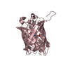
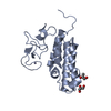
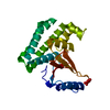
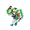
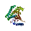
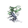
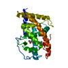
- PDB-3r7a: Crystal structure of phosphoglycerate mutase from Bacillus anthra... -
+
Open data
ID or keywords:
Loading...
-
Basic information
Entry
Database: PDB / ID: 3r7a
Title
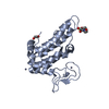
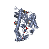
Crystal structure of phosphoglycerate mutase from Bacillus anthracis str. Sterne
Components
Phosphoglycerate mutase, putative
Keywords
TRANSFERASE / Structural Genomics / PSI-Biology / Midwest Center for Structural Genomics / MCSG / phosphoglycerate mutase / catalyzes the internal transfer of a phosphate group from C-3 to C-2 which results in the conversion of 3-phosphoglycerate (3PG) to 2-phosphoglycerate (2PG)
Function / homology
Function and homology information
regulation of pentose-phosphate shunt / fructose-2,6-bisphosphate 2-phosphatase activity / negative regulation of glycolytic process / cytosol Similarity search - Function
Resolution: 1.845→1.88 Å / Redundancy: 15.3 % / Rmerge(I) obs: 0.747 / Mean I/σ(I) obs: 4 / Num. unique all: 1967 / % possible all: 99.9
-
Processing
Software
Name
Version
Classification
NB
REFMAC
5.5.0109
refinement
PDB_EXTRACT
3.1
dataextraction
SBC-Collect
datacollection
HKL-3000
datareduction
HKL-3000
datascaling
HKL-3000
phasing
MLPHARE
phasing
DM
phasing
SHELXDE
phasing
RESOLVE
phasing
ARP/wARP
modelbuilding
Coot
modelbuilding
Refinement
Method to determine structure: SAD / Resolution: 1.845→71.725 Å / Cor.coef. Fo:Fc: 0.963 / Cor.coef. Fo:Fc free: 0.947 / Occupancy max: 1 / Occupancy min: 0.3 / SU B: 5.948 / SU ML: 0.082 / Cross valid method: THROUGHOUT / σ(F): 0 / ESU R: 0.412 / ESU R Free: 0.124 Stereochemistry target values: MAXIMUM LIKELIHOOD WITH PHASES Details: HYDROGENS HAVE BEEN ADDED IN THE RIDING POSITIONS U VALUES : WITH TLS ADDED
Rfactor
Num. reflection
% reflection
Selection details
Rfree
0.2043
2026
5 %
RANDOM
Rwork
0.1622
-
-
-
all
0.1643
40380
-
-
obs
0.1643
40380
99.91 %
-
Solvent computation
Ion probe radii: 0.8 Å / Shrinkage radii: 0.8 Å / VDW probe radii: 1.4 Å / Solvent model: MASK
In the structure databanks used in Yorodumi, some data are registered as the other names, "COVID-19 virus" and "2019-nCoV". Here are the details of the virus and the list of structure data.
Jan 31, 2019. EMDB accession codes are about to change! (news from PDBe EMDB page)
EMDB accession codes are about to change! (news from PDBe EMDB page)
The allocation of 4 digits for EMDB accession codes will soon come to an end. Whilst these codes will remain in use, new EMDB accession codes will include an additional digit and will expand incrementally as the available range of codes is exhausted. The current 4-digit format prefixed with “EMD-” (i.e. EMD-XXXX) will advance to a 5-digit format (i.e. EMD-XXXXX), and so on. It is currently estimated that the 4-digit codes will be depleted around Spring 2019, at which point the 5-digit format will come into force.
The EM Navigator/Yorodumi systems omit the EMD- prefix.
Related info.:Q: What is EMD? / ID/Accession-code notation in Yorodumi/EM Navigator
Yorodumi is a browser for structure data from EMDB, PDB, SASBDB, etc.
This page is also the successor to EM Navigator detail page, and also detail information page/front-end page for Omokage search.
The word "yorodu" (or yorozu) is an old Japanese word meaning "ten thousand". "mi" (miru) is to see.
Related info.:EMDB / PDB / SASBDB / Comparison of 3 databanks / Yorodumi Search / Aug 31, 2016. New EM Navigator & Yorodumi / Yorodumi Papers / Jmol/JSmol / Function and homology information / Changes in new EM Navigator and Yorodumi
 Movie
Movie Controller

 Yorodumi
 Open data
 Basic information
 Components
 Keywords
 Function and homology information
 X-RAY DIFFRACTION /
  Authors
 Citation
 Structure visualization
 Downloads & links
 Other downloads
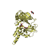

 PDBj
PDBj


 Assembly
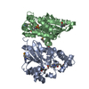


 Mass: 238.305 Da / Num. of mol.: 2 / Source method: obtained synthetically / Formula: C8H18N2O4S / Comment: pH buffer*YM
 Mass: 18.015 Da / Num. of mol.: 322 / Source method: isolated from a natural source / Formula: H2O
 Sample preparation
 / Beamline: 19-ID / Wavelength: 0.97912 Å
 Processing