 Movie
Movie Controller

[English] 日本語
 Yorodumi
- PDB-3r07: Structural analysis of an archaeal lipoylation system. A bi-parti... -
+ Open data

- Basic information
| Entry | Database: PDB / ID: 3r07 | ||||||
|---|---|---|---|---|---|---|---|
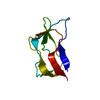
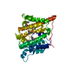
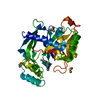
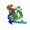
| Title | Structural analysis of an archaeal lipoylation system. A bi-partite lipoate protein ligase and its E2 lipoyl domain from Thermoplasma acidophilum | ||||||
 Components Components |
| ||||||
 Keywords Keywords | TRANSFERASE / Adenylate-forming enzyme / Ligase / bi-partite / ATP-binding | ||||||
| Function / homology |  Function and homology information Function and homology informationlipoate-protein ligase / lipoate-protein ligase activity / lipoic acid binding / protein lipoylation / protein-containing complex / ATP binding / metal ion binding / cytoplasm Similarity search - Function | ||||||
| Biological species |   Thermoplasma acidophilum DSM 1728 (acidophilic) Thermoplasma acidophilum DSM 1728 (acidophilic) | ||||||
| Method |  X-RAY DIFFRACTION / SYNCHROTRON / MOLECULAR REPLACEMENT / Resolution: 2.7 Å X-RAY DIFFRACTION / SYNCHROTRON / MOLECULAR REPLACEMENT / Resolution: 2.7 Å | ||||||
 Authors Authors | Posner, M.G. / Upadhyay, U. / Crennell, S. | ||||||
 Citation Citation | Journal: To be Published Title: Structural analysis of an archaeal lipoylation system. A bi-partite lipoate protein ligase and its E2 lipoyl domain from Thermoplasma acidophilum Authors: Posner, M.G. / Upadhyay, A. / Crennell, S. / Danson, M.J. / Bagby, S. | ||||||
| History |
|
- Structure visualization
| Structure viewer | Molecule: MolmilJmol/JSmol |
|---|
- Downloads & links
-Download
| PDBx/mmCIF format | 3r07.cif.gz | 85.3 KB | Display | PDBx/mmCIF format |
|---|---|---|---|---|
| PDB format | pdb3r07.ent.gz | 64.4 KB | Display | PDB format |
| PDBx/mmJSON format | 3r07.json.gz | Tree view | PDBx/mmJSON format | |
| Others |  Other downloads Other downloads |
-Validation report
| Arichive directory | https://data.pdbj.org/pub/pdb/validation_reports/r0/3r07ftp://data.pdbj.org/pub/pdb/validation_reports/r0/3r07 | HTTPS FTP |
|---|
-Related structure data
| Related structure data |  2arsS S: Starting model for refinement |
|---|---|
| Similar structure data |


-Links
 PDBj
PDBj
- Assembly
| Deposited unit | 
| ||||||||
|---|---|---|---|---|---|---|---|---|---|
| 1 |
| ||||||||
| Unit cell |
|
-Components
| #1: Protein | Mass: 32695.148 Da / Num. of mol.: 1 Source method: isolated from a genetically manipulated source Details: Co-expression Source: (gene. exp.) Thermoplasma acidophilum DSM 1728 (acidophilic)Strain: ATCC 25905 / DSM 1728 / JCM 9062 / NBRC 15155 / AMRC-C165 Gene: lplA, Ta0514 / Plasmid: pET19b / Production host:  | ||||
|---|---|---|---|---|---|
| #2: Protein | Mass: 10561.207 Da / Num. of mol.: 1 Source method: isolated from a genetically manipulated source Details: Co-expression Source: (gene. exp.) Thermoplasma acidophilum DSM 1728 (acidophilic)Strain: ATCC 25905 / DSM 1728 / JCM 9062 / NBRC 15155 / AMRC-C165 Gene: Ta0513, Ta0513m / Plasmid: pET28a / Production host: | ||||
| #3: Chemical |   Mass: 118.174 Da / Num. of mol.: 2 / Source method: obtained synthetically / Formula: C6H14O2 / Comment: precipitant*YM Mass: 118.174 Da / Num. of mol.: 2 / Source method: obtained synthetically / Formula: C6H14O2 / Comment: precipitant*YM#4: Chemical |   Mass: 59.044 Da / Num. of mol.: 2 / Source method: obtained synthetically / Formula: C2H3O2 Mass: 59.044 Da / Num. of mol.: 2 / Source method: obtained synthetically / Formula: C2H3O2#5: Water | ChemComp-HOH / |  Mass: 18.015 Da / Num. of mol.: 47 / Source method: isolated from a natural source / Formula: H2O Mass: 18.015 Da / Num. of mol.: 47 / Source method: isolated from a natural source / Formula: H2O |
-Experimental details
-Experiment
| Experiment | Method: X-RAY DIFFRACTION / Number of used crystals: 1 |
|---|
- Sample preparation
| Crystal | Density Matthews: 3.42 Å3/Da / Density % sol: 64.04 % |
|---|---|
| Crystal grow | Temperature: 291 K / pH: 4.6 Details: 40% v/v MPD, 0.1M sodium acetate, 0.02M calcium chloride, pH 4.6, VAPOR DIFFUSION, HANGING DROP, temperature 291K |
-Data collection
| Diffraction | Mean temperature: 200 K |
|---|---|
| Diffraction source | Source: SYNCHROTRON / Site: Diamond  / Beamline: I02 / Wavelength: 0.9795 / Beamline: I02 / Wavelength: 0.9795 |
| Detector | Type: ADSC QUANTUM 315r / Detector: CCD / Date: Oct 26, 2009 / Details: VORTEX FLUORESCENCE DETECTOR |
| Radiation | Protocol: SINGLE WAVELENGTH / Monochromatic (M) / Laue (L): M / Scattering type: x-ray |
| Radiation wavelength | Wavelength: 0.9795 Å / Relative weight: 1 |
| Reflection | Resolution: 2.7→38.81 Å |
- Processing
| Software |
| ||||||||||||||||||||||||||||||||||||||||||||||||||||||||||||||||||||||||||||||||||||||||||||||||||||||||||||||||||||||||||||||||||||||||||||||||||||||||||||||||||||||||||
|---|---|---|---|---|---|---|---|---|---|---|---|---|---|---|---|---|---|---|---|---|---|---|---|---|---|---|---|---|---|---|---|---|---|---|---|---|---|---|---|---|---|---|---|---|---|---|---|---|---|---|---|---|---|---|---|---|---|---|---|---|---|---|---|---|---|---|---|---|---|---|---|---|---|---|---|---|---|---|---|---|---|---|---|---|---|---|---|---|---|---|---|---|---|---|---|---|---|---|---|---|---|---|---|---|---|---|---|---|---|---|---|---|---|---|---|---|---|---|---|---|---|---|---|---|---|---|---|---|---|---|---|---|---|---|---|---|---|---|---|---|---|---|---|---|---|---|---|---|---|---|---|---|---|---|---|---|---|---|---|---|---|---|---|---|---|---|---|---|---|---|---|
| Refinement | Method to determine structure: MOLECULAR REPLACEMENT Starting model: 2ARS Resolution: 2.7→38.81 Å / Cor.coef. Fo:Fc: 0.934 / Cor.coef. Fo:Fc free: 0.892 / SU B: 9.005 / SU ML: 0.192 / Cross valid method: THROUGHOUT / ESU R Free: 0.305 / Stereochemistry target values: MAXIMUM LIKELIHOOD / Details: HYDROGENS HAVE BEEN ADDED IN THE RIDING POSITIONS
| ||||||||||||||||||||||||||||||||||||||||||||||||||||||||||||||||||||||||||||||||||||||||||||||||||||||||||||||||||||||||||||||||||||||||||||||||||||||||||||||||||||||||||
| Solvent computation | Ion probe radii: 0.8 Å / Shrinkage radii: 0.8 Å / VDW probe radii: 1.2 Å / Solvent model: MASK | ||||||||||||||||||||||||||||||||||||||||||||||||||||||||||||||||||||||||||||||||||||||||||||||||||||||||||||||||||||||||||||||||||||||||||||||||||||||||||||||||||||||||||
| Displacement parameters | Biso mean: 51.56 Å2
| ||||||||||||||||||||||||||||||||||||||||||||||||||||||||||||||||||||||||||||||||||||||||||||||||||||||||||||||||||||||||||||||||||||||||||||||||||||||||||||||||||||||||||
| Refinement step | Cycle: LAST / Resolution: 2.7→38.81 Å
| ||||||||||||||||||||||||||||||||||||||||||||||||||||||||||||||||||||||||||||||||||||||||||||||||||||||||||||||||||||||||||||||||||||||||||||||||||||||||||||||||||||||||||
| Refine LS restraints |
| ||||||||||||||||||||||||||||||||||||||||||||||||||||||||||||||||||||||||||||||||||||||||||||||||||||||||||||||||||||||||||||||||||||||||||||||||||||||||||||||||||||||||||
| LS refinement shell | Resolution: 2.7→2.77 Å / Total num. of bins used: 20
|
