 Movie
Movie Controller

[English] 日本語
 Yorodumi
- PDB-3qs2: Crystal structure of the biofilm forming subunit of the E. coli c... -
+ Open data

- Basic information
| Entry | Database: PDB / ID: 3qs2 | ||||||
|---|---|---|---|---|---|---|---|
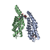
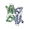
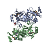
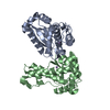
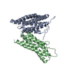
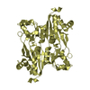
| Title | Crystal structure of the biofilm forming subunit of the E. coli common pilus: full length domain swapped dimer of EcpA | ||||||
 Components Components | Fimbrillin matB homolog | ||||||
 Keywords Keywords | CELL ADHESION / pilin / Ig-like fold / biofilms / adhesion / Immunoglobulin-like fold / major pilin domain involved in biofilms / intermolecular and hydrophobic abiotic surface binding / extracellular membrane | ||||||
| Function / homology |  Function and homology information Function and homology information | ||||||
| Biological species |  | ||||||
| Method |  X-RAY DIFFRACTION / SYNCHROTRON / SAD / Resolution: 1.78 Å X-RAY DIFFRACTION / SYNCHROTRON / SAD / Resolution: 1.78 Å | ||||||
 Authors Authors | Garnett, J.A. / Matthews, S.J. | ||||||
 Citation Citation | Journal: Proc.Natl.Acad.Sci.USA / Year: 2012 Title: Structural insights into the biogenesis and biofilm formation by the Escherichia coli common pilus. Authors: Garnett, J.A. / Martinez-Santos, V.I. / Saldana, Z. / Pape, T. / Hawthorne, W. / Chan, J. / Simpson, P.J. / Cota, E. / Puente, J.L. / Giron, J.A. / Matthews, S. | ||||||
| History |
|
- Structure visualization
| Structure viewer | Molecule: MolmilJmol/JSmol |
|---|
- Downloads & links
-Download
| PDBx/mmCIF format | 3qs2.cif.gz | 148.7 KB | Display | PDBx/mmCIF format |
|---|---|---|---|---|
| PDB format | pdb3qs2.ent.gz | 117.6 KB | Display | PDB format |
| PDBx/mmJSON format | 3qs2.json.gz | Tree view | PDBx/mmJSON format | |
| Others |  Other downloads Other downloads |
-Validation report
| Arichive directory | https://data.pdbj.org/pub/pdb/validation_reports/qs/3qs2ftp://data.pdbj.org/pub/pdb/validation_reports/qs/3qs2 | HTTPS FTP |
|---|
-Related structure data


-Links
 PDBj
PDBj- Assembly
| Deposited unit | 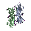
| ||||||||
|---|---|---|---|---|---|---|---|---|---|
| 1 |
| ||||||||
| Unit cell |
|
-Components
| #1: Protein | Mass: 19742.523 Da / Num. of mol.: 2 / Fragment: Full length processed EcpA, rsidues 23-195 Source method: isolated from a genetically manipulated source Source: (gene. exp.) #2: Chemical | ChemComp-IOD /   Mass: 126.904 Da / Num. of mol.: 21 / Source method: obtained synthetically / Formula: I Mass: 126.904 Da / Num. of mol.: 21 / Source method: obtained synthetically / Formula: I#3: Water | ChemComp-HOH / |  Mass: 18.015 Da / Num. of mol.: 460 / Source method: isolated from a natural source / Formula: H2O Mass: 18.015 Da / Num. of mol.: 460 / Source method: isolated from a natural source / Formula: H2O |
|---|
-Experimental details
-Experiment
| Experiment | Method: X-RAY DIFFRACTION / Number of used crystals: 1 |
|---|
- Sample preparation
| Crystal | Density Matthews: 2.32 Å3/Da / Density % sol: 46.9 % |
|---|---|
| Crystal grow | Temperature: 293 K / pH: 3.2 Details: 15% PEG 3350, 100 mM phosphate/citrate, 200 mM NaI, pH 3.2, VAPOR DIFFUSION, SITTING DROP, temperature 293K |
-Data collection
| Diffraction | Mean temperature: 100 K |
|---|---|
| Diffraction source | Source: SYNCHROTRON / Site: Diamond  / Beamline: I04 / Wavelength: 1.54 / Beamline: I04 / Wavelength: 1.54 |
| Detector | Type: ADSC QUANTUM 315r / Detector: CCD / Date: Sep 24, 2010 / Details: MIRRORS |
| Radiation | Monochromator: SI (111) DOUBLE CRYSTAL / Protocol: SINGLE WAVELENGTH / Monochromatic (M) / Laue (L): M / Scattering type: x-ray |
| Radiation wavelength | Wavelength: 1.54 Å / Relative weight: 1 |
| Reflection | Resolution: 1.78→60.01 Å / Num. obs: 35831 / % possible obs: 100 % / Observed criterion σ(I): 2 / Redundancy: 26.5 % / Rsym value: 0.35 / Net I/σ(I): 3.7 |
| Reflection shell | Resolution: 1.78→1.88 Å / Redundancy: 26.4 % / Mean I/σ(I) obs: 2.1 / Rsym value: 0.11 / % possible all: 100 |
- Processing
| Software |
| ||||||||||||||||||||||||||||||||||||||||||||||||||||||||||||||||||||||||||||||||||||||||||||||||||||||||||||||||||||||||||||||||||||||||||||||||||||||||||||||||||||||||||
|---|---|---|---|---|---|---|---|---|---|---|---|---|---|---|---|---|---|---|---|---|---|---|---|---|---|---|---|---|---|---|---|---|---|---|---|---|---|---|---|---|---|---|---|---|---|---|---|---|---|---|---|---|---|---|---|---|---|---|---|---|---|---|---|---|---|---|---|---|---|---|---|---|---|---|---|---|---|---|---|---|---|---|---|---|---|---|---|---|---|---|---|---|---|---|---|---|---|---|---|---|---|---|---|---|---|---|---|---|---|---|---|---|---|---|---|---|---|---|---|---|---|---|---|---|---|---|---|---|---|---|---|---|---|---|---|---|---|---|---|---|---|---|---|---|---|---|---|---|---|---|---|---|---|---|---|---|---|---|---|---|---|---|---|---|---|---|---|---|---|---|---|
| Refinement | Method to determine structure: SAD / Resolution: 1.78→60.01 Å / Cor.coef. Fo:Fc: 0.952 / Cor.coef. Fo:Fc free: 0.932 / SU B: 3.96 / SU ML: 0.073 / Isotropic thermal model: TLS / Cross valid method: THROUGHOUT / ESU R Free: 0.125 / Stereochemistry target values: MAXIMUM LIKELIHOOD / Details: HYDROGENS HAVE BEEN USED IF PRESENT IN THE INPUT
| ||||||||||||||||||||||||||||||||||||||||||||||||||||||||||||||||||||||||||||||||||||||||||||||||||||||||||||||||||||||||||||||||||||||||||||||||||||||||||||||||||||||||||
| Solvent computation | Ion probe radii: 0.8 Å / Shrinkage radii: 0.8 Å / VDW probe radii: 1.2 Å / Solvent model: MASK | ||||||||||||||||||||||||||||||||||||||||||||||||||||||||||||||||||||||||||||||||||||||||||||||||||||||||||||||||||||||||||||||||||||||||||||||||||||||||||||||||||||||||||
| Displacement parameters | Biso mean: 16.81 Å2
| ||||||||||||||||||||||||||||||||||||||||||||||||||||||||||||||||||||||||||||||||||||||||||||||||||||||||||||||||||||||||||||||||||||||||||||||||||||||||||||||||||||||||||
| Refinement step | Cycle: LAST / Resolution: 1.78→60.01 Å
| ||||||||||||||||||||||||||||||||||||||||||||||||||||||||||||||||||||||||||||||||||||||||||||||||||||||||||||||||||||||||||||||||||||||||||||||||||||||||||||||||||||||||||
| Refine LS restraints |
| ||||||||||||||||||||||||||||||||||||||||||||||||||||||||||||||||||||||||||||||||||||||||||||||||||||||||||||||||||||||||||||||||||||||||||||||||||||||||||||||||||||||||||
| LS refinement shell | Resolution: 1.78→1.83 Å / Total num. of bins used: 20
| ||||||||||||||||||||||||||||||||||||||||||||||||||||||||||||||||||||||||||||||||||||||||||||||||||||||||||||||||||||||||||||||||||||||||||||||||||||||||||||||||||||||||||
| Refinement TLS params. | Method: refined / Refine-ID: X-RAY DIFFRACTION
| ||||||||||||||||||||||||||||||||||||||||||||||||||||||||||||||||||||||||||||||||||||||||||||||||||||||||||||||||||||||||||||||||||||||||||||||||||||||||||||||||||||||||||
| Refinement TLS group |
|
