 Movie
Movie Controller

[English] 日本語
 Yorodumi
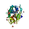
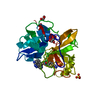
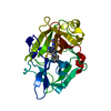
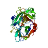
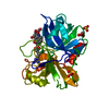
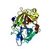
- PDB-3qn7: Potent and selective bicyclic peptide inhibitor (UK18) of human u... -
+ Open data

- Basic information
| Entry | Database: PDB / ID: 3qn7 | ||||||
|---|---|---|---|---|---|---|---|
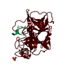
| Title | Potent and selective bicyclic peptide inhibitor (UK18) of human urokinase-type plasminogen activator(uPA) | ||||||
 Components Components |
| ||||||
 Keywords Keywords | HYDROLASE/HYDROLASE INHIBITOR / Bicyclic peptide inhibitor / Chymotrypsin fold / Serine protease / Urokinase receptor (uPAR) / Extracellular / HYDROLASE-HYDROLASE INHIBITOR complex | ||||||
| Function / homology |  Function and homology information Function and homology informationu-plasminogen activator / regulation of smooth muscle cell-matrix adhesion / urokinase plasminogen activator signaling pathway / regulation of plasminogen activation / regulation of integrin-mediated signaling pathway / protein complex involved in cell-matrix adhesion / regulation of fibrinolysis / regulation of wound healing / negative regulation of plasminogen activation / serine-type endopeptidase complex ...u-plasminogen activator / regulation of smooth muscle cell-matrix adhesion / urokinase plasminogen activator signaling pathway / regulation of plasminogen activation / regulation of integrin-mediated signaling pathway / protein complex involved in cell-matrix adhesion / regulation of fibrinolysis / regulation of wound healing / negative regulation of plasminogen activation / serine-type endopeptidase complex / regulation of smooth muscle cell migration / Dissolution of Fibrin Clot / regulation of cell adhesion mediated by integrin / smooth muscle cell migration / plasminogen activation / tertiary granule membrane / negative regulation of fibrinolysis / regulation of cell adhesion / serine protease inhibitor complex / specific granule membrane / fibrinolysis / positive regulation of epidermal growth factor receptor signaling pathway / chemotaxis / blood coagulation / regulation of cell population proliferation / response to hypoxia / positive regulation of cell migration / receptor ligand activity / serine-type endopeptidase activity / external side of plasma membrane / focal adhesion / Neutrophil degranulation / cell surface / signal transduction / proteolysis / : / extracellular exosome / extracellular region / plasma membrane Similarity search - Function | ||||||
| Biological species |  Homo sapiens (human) Homo sapiens (human) | ||||||
| Method |  X-RAY DIFFRACTION / SYNCHROTRON / MOLECULAR REPLACEMENT / Resolution: 1.9 Å X-RAY DIFFRACTION / SYNCHROTRON / MOLECULAR REPLACEMENT / Resolution: 1.9 Å | ||||||
 Authors Authors | Angelini, A. / Cendron, L. / Touati, J. / Winter, G. / Zanotti, G. / Heinis, C. | ||||||
 Citation Citation | Journal: Acs Chem.Biol. / Year: 2012 Title: Bicyclic peptide inhibitor reveals large contact interface with a protease target Authors: Angelini, A. / Cendron, L. / Chen, S. / Touati, J. / Winter, G. / Zanotti, G. / Heinis, C. #1: Journal: J.Struct.Biol. / Year: 2007Title: Structural basis of specificity of a peptidyl urokinase inhibitor, upain-1 Authors: Zhao, G. / Yuan, C. / Wind, T. / Huang, Z. / Andreasen, P.A. / Huang, M. #2: Journal: Structure / Year: 1995Title: The crystal structure of the catalytic domain of human urokinase-type plasminogen activator Authors: Spraggon, G. / Phillips, C. / Nowak, U.K. / Ponting, C.P. / Saunders, D. / Dobson, C.M. / Stuart, D.I. / Jones, E.Y. | ||||||
| History |
|
- Structure visualization
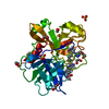
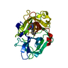
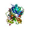
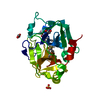
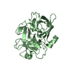
| Structure viewer | Molecule: MolmilJmol/JSmol |
|---|
- Downloads & links
-Download
| PDBx/mmCIF format | 3qn7.cif.gz | 69.5 KB | Display | PDBx/mmCIF format |
|---|---|---|---|---|
| PDB format | pdb3qn7.ent.gz | 51.1 KB | Display | PDB format |
| PDBx/mmJSON format | 3qn7.json.gz | Tree view | PDBx/mmJSON format | |
| Others |  Other downloads Other downloads |
-Validation report
| Arichive directory | https://data.pdbj.org/pub/pdb/validation_reports/qn/3qn7ftp://data.pdbj.org/pub/pdb/validation_reports/qn/3qn7 | HTTPS FTP |
|---|
-Related structure data
| Related structure data |  2nwnS S: Starting model for refinement |
|---|---|
| Similar structure data |


-Links
 PDBj
PDBj

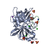
- Assembly
| Deposited unit | 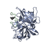
| ||||||||
|---|---|---|---|---|---|---|---|---|---|
| 1 |
| ||||||||
| Unit cell |
|
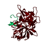
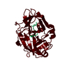
-Components
| #1: Protein | Mass: 28442.373 Da / Num. of mol.: 1 Fragment: Catalytic domain, Urokinase-type plasminogen activator chain B Mutation: C122A, N145Q Source method: isolated from a genetically manipulated source Source: (gene. exp.) Homo sapiens (human) / Gene: PLAU / Plasmid: pSecTagA / Cell (production host): mammalian cellsCell line (production host): Human embryonic kidney cells (HEK-293) Production host: Homo sapiens (human) / References: UniProt: P00749, u-plasminogen activator |
|---|---|
| #2: Protein/peptide | Mass: 1792.998 Da / Num. of mol.: 1 / Source method: obtained synthetically / Details: Chemical synthesis |
| #3: Chemical | ChemComp-ZBR /   Mass: 356.880 Da / Num. of mol.: 1 / Source method: obtained synthetically / Formula: C9H9Br3 Mass: 356.880 Da / Num. of mol.: 1 / Source method: obtained synthetically / Formula: C9H9Br3 |
| #4: Water | ChemComp-HOH /  Mass: 18.015 Da / Num. of mol.: 132 / Source method: isolated from a natural source / Formula: H2O Mass: 18.015 Da / Num. of mol.: 132 / Source method: isolated from a natural source / Formula: H2O |
| Has protein modification | Y |
-Experimental details
-Experiment
| Experiment | Method: X-RAY DIFFRACTION / Number of used crystals: 1 |
|---|
- Sample preparation
| Crystal | Density Matthews: 2 Å3/Da / Density % sol: 38.46 % |
|---|---|
| Crystal grow | Temperature: 293 K / Method: vapor diffusion, hanging drop / pH: 4.3 Details: 2M ammonium sulfate, 0.05M sodium citrate, 5%(v/v) PEG 400, pH 4.3, VAPOR DIFFUSION, HANGING DROP, temperature 293K |
-Data collection
| Diffraction | Mean temperature: 100 K |
|---|---|
| Diffraction source | Source: SYNCHROTRON / Site: ESRF  / Beamline: ID14-4 / Wavelength: 0.939 Å / Beamline: ID14-4 / Wavelength: 0.939 Å |
| Detector | Type: ADSC QUANTUM 315r / Detector: CCD / Date: Oct 29, 2010 / Details: Toroidal focusing mirrors |
| Radiation | Protocol: SINGLE WAVELENGTH / Monochromatic (M) / Laue (L): M / Scattering type: x-ray |
| Radiation wavelength | Wavelength: 0.939 Å / Relative weight: 1 |
| Reflection | Resolution: 1.9→39.57 Å / Num. all: 18155 / Num. obs: 18155 / % possible obs: 99.8 % / Observed criterion σ(F): 0 / Observed criterion σ(I): 0 / Redundancy: 5.4 % / Rmerge(I) obs: 0.079 / Net I/σ(I): 12.2 |
| Reflection shell | Resolution: 1.9→2 Å / Redundancy: 5.4 % / Rmerge(I) obs: 0.405 / Mean I/σ(I) obs: 3.9 / % possible all: 100 |
- Processing
| Software |
| ||||||||||||||||||||||||||||||||||||||||||||||||||||||||||||||||||||||||||||||||||||||||||||||||||||||||||||||||||||||||||||||||||||||||||||||||||||||||||||||||||||||||||
|---|---|---|---|---|---|---|---|---|---|---|---|---|---|---|---|---|---|---|---|---|---|---|---|---|---|---|---|---|---|---|---|---|---|---|---|---|---|---|---|---|---|---|---|---|---|---|---|---|---|---|---|---|---|---|---|---|---|---|---|---|---|---|---|---|---|---|---|---|---|---|---|---|---|---|---|---|---|---|---|---|---|---|---|---|---|---|---|---|---|---|---|---|---|---|---|---|---|---|---|---|---|---|---|---|---|---|---|---|---|---|---|---|---|---|---|---|---|---|---|---|---|---|---|---|---|---|---|---|---|---|---|---|---|---|---|---|---|---|---|---|---|---|---|---|---|---|---|---|---|---|---|---|---|---|---|---|---|---|---|---|---|---|---|---|---|---|---|---|---|---|---|
| Refinement | Method to determine structure: MOLECULAR REPLACEMENT Starting model: 2NWN.pdb Resolution: 1.9→39.57 Å / Cor.coef. Fo:Fc: 0.954 / Cor.coef. Fo:Fc free: 0.922 / SU B: 4.495 / SU ML: 0.134 / Cross valid method: THROUGHOUT / σ(F): 0 / σ(I): 0 / ESU R Free: 0.173 / Stereochemistry target values: MAXIMUM LIKELIHOOD / Details: HYDROGENS HAVE BEEN ADDED IN THE RIDING POSITIONS
| ||||||||||||||||||||||||||||||||||||||||||||||||||||||||||||||||||||||||||||||||||||||||||||||||||||||||||||||||||||||||||||||||||||||||||||||||||||||||||||||||||||||||||
| Solvent computation | Ion probe radii: 0.8 Å / Shrinkage radii: 0.8 Å / VDW probe radii: 1.4 Å / Solvent model: BABINET MODEL WITH MASK | ||||||||||||||||||||||||||||||||||||||||||||||||||||||||||||||||||||||||||||||||||||||||||||||||||||||||||||||||||||||||||||||||||||||||||||||||||||||||||||||||||||||||||
| Displacement parameters | Biso mean: 34.287 Å2
| ||||||||||||||||||||||||||||||||||||||||||||||||||||||||||||||||||||||||||||||||||||||||||||||||||||||||||||||||||||||||||||||||||||||||||||||||||||||||||||||||||||||||||
| Refinement step | Cycle: LAST / Resolution: 1.9→39.57 Å
| ||||||||||||||||||||||||||||||||||||||||||||||||||||||||||||||||||||||||||||||||||||||||||||||||||||||||||||||||||||||||||||||||||||||||||||||||||||||||||||||||||||||||||
| Refine LS restraints |
| ||||||||||||||||||||||||||||||||||||||||||||||||||||||||||||||||||||||||||||||||||||||||||||||||||||||||||||||||||||||||||||||||||||||||||||||||||||||||||||||||||||||||||
| LS refinement shell | Resolution: 1.9→1.95 Å / Total num. of bins used: 20
|
