 Movie
Movie Controller

[English] 日本語
 Yorodumi
- PDB-3qjk: Structure of Calcium Binding Protein-1 from Entamoeba histolytica... -
+ Open data

- Basic information
| Entry | Database: PDB / ID: 3qjk | ||||||
|---|---|---|---|---|---|---|---|
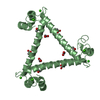
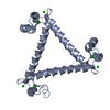
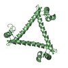
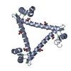
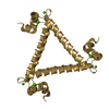
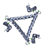
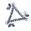
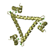
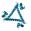
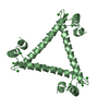
| Title | Structure of Calcium Binding Protein-1 from Entamoeba histolytica in complex with Lead | ||||||
 Components Components | Calcium-binding protein | ||||||
 Keywords Keywords | METAL BINDING PROTEIN / EF-hand | ||||||
| Function / homology |  Function and homology information Function and homology informationpositive regulation of macropinocytosis / regulation of actin filament bundle assembly / regulation of protein kinase activity / pseudopodium / actin monomer binding / phagocytosis / phagocytic cup / positive regulation of phagocytosis / actin filament binding / calcium ion binding / cytoplasm Similarity search - Function | ||||||
| Biological species |   Entamoeba histolytica (eukaryote) Entamoeba histolytica (eukaryote) | ||||||
| Method |  X-RAY DIFFRACTION / MOLECULAR REPLACEMENT / Resolution: 3.001 Å X-RAY DIFFRACTION / MOLECULAR REPLACEMENT / Resolution: 3.001 Å | ||||||
 Authors Authors | Kumar, S. / Gourinath, S. | ||||||
 Citation Citation | Journal: To be Published Title: Structure of Calcium Binding Protein-1 from Entamoeba histolytica in complex with Lead Authors: Kumar, S. / Gourinath, S. | ||||||
| History |
|
- Structure visualization
| Structure viewer | Molecule: MolmilJmol/JSmol |
|---|
- Downloads & links
-Download
| PDBx/mmCIF format | 3qjk.cif.gz | 40.7 KB | Display | PDBx/mmCIF format |
|---|---|---|---|---|
| PDB format | pdb3qjk.ent.gz | 27.1 KB | Display | PDB format |
| PDBx/mmJSON format | 3qjk.json.gz | Tree view | PDBx/mmJSON format | |
| Others |  Other downloads Other downloads |
-Validation report
| Summary document | 3qjk_validation.pdf.gz | 447.9 KB | Display | wwPDB validaton report |
|---|---|---|---|---|
| Full document | 3qjk_full_validation.pdf.gz | 448 KB | Display | |
| Data in XML | 3qjk_validation.xml.gz | 7 KB | Display | |
| Data in CIF | 3qjk_validation.cif.gz | 8.7 KB | Display | |
| Arichive directory | https://data.pdbj.org/pub/pdb/validation_reports/qj/3qjkftp://data.pdbj.org/pub/pdb/validation_reports/qj/3qjk | HTTPS FTP |
-Related structure data
| Related structure data |  2nxqS S: Starting model for refinement |
|---|---|
| Similar structure data |
-Links
 PDBj
PDBj


- Assembly
| Deposited unit | 
| ||||||||
|---|---|---|---|---|---|---|---|---|---|
| 1 | 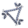
| ||||||||
| 2 | 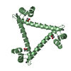
| ||||||||
| Unit cell |
|
-Components
| #1: Protein | Mass: 14970.852 Da / Num. of mol.: 2 Source method: isolated from a genetically manipulated source Details: cytosol / Source: (gene. exp.) Entamoeba histolytica (eukaryote) / Strain: HM1:IMSS / Gene: EhCaBP1 / Plasmid: pET-3c / Production host:  #2: Chemical | ChemComp-PB /   Mass: 207.200 Da / Num. of mol.: 4 / Source method: obtained synthetically / Formula: Pb Mass: 207.200 Da / Num. of mol.: 4 / Source method: obtained synthetically / Formula: Pb#3: Chemical |   Mass: 59.044 Da / Num. of mol.: 2 / Source method: obtained synthetically / Formula: C2H3O2 Mass: 59.044 Da / Num. of mol.: 2 / Source method: obtained synthetically / Formula: C2H3O2#4: Water | ChemComp-HOH / |  Mass: 18.015 Da / Num. of mol.: 17 / Source method: isolated from a natural source / Formula: H2O Mass: 18.015 Da / Num. of mol.: 17 / Source method: isolated from a natural source / Formula: H2O |
|---|
-Experimental details
-Experiment
| Experiment | Method: X-RAY DIFFRACTION / Number of used crystals: 1 |
|---|
- Sample preparation
| Crystal | Density Matthews: 2.83 Å3/Da / Density % sol: 56.48 % |
|---|---|
| Crystal grow | Temperature: 289 K / Method: vapor diffusion, hanging drop / pH: 3.6 Details: 60-65% MPD, 50mM NaOAc, 5mM Pb(NO3)2, pH 3.6, VAPOR DIFFUSION, HANGING DROP, temperature 289K |
-Data collection
| Diffraction | Mean temperature: 100 K |
|---|---|
| Diffraction source | Source: ROTATING ANODE / Type: BRUKER AXS MICROSTAR |
| Detector | Type: MAR scanner 345 mm plate / Detector: IMAGE PLATE / Date: Jun 5, 2008 / Details: mirrors |
| Radiation | Monochromator: GRAPHITE / Protocol: SINGLE WAVELENGTH / Monochromatic (M) / Laue (L): M / Scattering type: x-ray |
| Radiation wavelength | Relative weight: 1 |
| Reflection | Resolution: 3→30 Å / Num. all: 6777 / Num. obs: 6763 / % possible obs: 87 % / Observed criterion σ(F): 0 / Observed criterion σ(I): 0 / Redundancy: 2.9 % / Rmerge(I) obs: 0.054 / Rsym value: 0.0364 / Net I/σ(I): 8.8 |
| Reflection shell | Resolution: 3→3.11 Å / Redundancy: 2.9 % / Rmerge(I) obs: 0.054 / Mean I/σ(I) obs: 1.1 / Rsym value: 0.364 / % possible all: 99.7 |
- Processing
| Software |
| ||||||||||||||||||||||||||||||||||||
|---|---|---|---|---|---|---|---|---|---|---|---|---|---|---|---|---|---|---|---|---|---|---|---|---|---|---|---|---|---|---|---|---|---|---|---|---|---|
| Refinement | Method to determine structure: MOLECULAR REPLACEMENT Starting model: PDB ENTRY 2NXQ Resolution: 3.001→28.082 Å / Occupancy max: 1 / Occupancy min: 1 / SU ML: 0.5 / Isotropic thermal model: RESTRAINED / σ(F): 1.39 / Phase error: 29.01 / Stereochemistry target values: ML / Details: BULK SOLVENT MODEL USED
| ||||||||||||||||||||||||||||||||||||
| Solvent computation | Shrinkage radii: 0.86 Å / VDW probe radii: 1.1 Å / Solvent model: FLAT BULK SOLVENT MODEL / Bsol: 57.955 Å2 / ksol: 0.312 e/Å3 | ||||||||||||||||||||||||||||||||||||
| Displacement parameters | Biso mean: 75.1336 Å2
| ||||||||||||||||||||||||||||||||||||
| Refine analyze |
| ||||||||||||||||||||||||||||||||||||
| Refinement step | Cycle: LAST / Resolution: 3.001→28.082 Å
| ||||||||||||||||||||||||||||||||||||
| Refine LS restraints |
| ||||||||||||||||||||||||||||||||||||
| LS refinement shell | Refine-ID: X-RAY DIFFRACTION / Total num. of bins used: 5
|
