Type: MAR CCD 165 mm / Detector: CCD / Date: Nov 5, 2010
Radiation
Protocol: SINGLE WAVELENGTH / Monochromatic (M) / Laue (L): M / Scattering type: x-ray
Radiation wavelength
Wavelength: 0.979981 Å / Relative weight: 1
Reflection
Resolution: 2→50 Å / Num. obs: 43206 / % possible obs: 99.4 % / Observed criterion σ(I): 2 / Redundancy: 4.53 % / Biso Wilson estimate: 33.334 Å2 / Rmerge(I) obs: 0.026 / Net I/σ(I): 36.51
Reflection shell
Resolution: 2→2.05 Å / Redundancy: 4.2 % / Rmerge(I) obs: 0.179 / Mean I/σ(I) obs: 7.39 / Num. unique all: 3103 / % possible all: 97.9
-
Processing
Software
Name
Version
Classification
BEST
datacollection
Auto-Rickshaw
phasing
REFMAC
5.5.0109
refinement
XDS
datareduction
XSCALE
datascaling
Refinement
Method to determine structure: SAD / Resolution: 2→19.57 Å / Cor.coef. Fo:Fc: 0.942 / Cor.coef. Fo:Fc free: 0.931 / SU B: 7.663 / SU ML: 0.102 / Cross valid method: THROUGHOUT / σ(I): 2 / ESU R Free: 0.143 / Stereochemistry target values: MAXIMUM LIKELIHOOD / Details: HYDROGENS HAVE BEEN ADDED IN THE RIDING POSITIONS
Rfactor
Num. reflection
% reflection
Selection details
Rfree
0.2277
2174
5 %
RANDOM
Rwork
0.2036
-
-
-
obs
0.2048
41031
99.42 %
-
all
-
43206
-
-
Solvent computation
Ion probe radii: 0.8 Å / Shrinkage radii: 0.8 Å / VDW probe radii: 1.4 Å / Solvent model: MASK
Displacement parameters
Biso mean: 35.4208 Å2
Baniso -1
Baniso -2
Baniso -3
1-
0.54 Å2
0 Å2
0.64 Å2
2-
-
1.12 Å2
0 Å2
3-
-
-
-1.24 Å2
Refinement step
Cycle: LAST / Resolution: 2→19.57 Å
Protein
Nucleic acid
Ligand
Solvent
Total
Num. atoms
3489
0
17
296
3802
Refine LS restraints
Refine-ID
Type
Dev ideal
Dev ideal target
Number
X-RAY DIFFRACTION
r_bond_refined_d
0.008
0.022
3655
X-RAY DIFFRACTION
r_angle_refined_deg
1.026
1.995
4970
X-RAY DIFFRACTION
r_dihedral_angle_1_deg
4.857
5
454
X-RAY DIFFRACTION
r_dihedral_angle_2_deg
38.101
26.438
160
X-RAY DIFFRACTION
r_dihedral_angle_3_deg
12.876
15
686
X-RAY DIFFRACTION
r_dihedral_angle_4_deg
7.786
15
4
X-RAY DIFFRACTION
r_chiral_restr
0.074
0.2
598
X-RAY DIFFRACTION
r_gen_planes_refined
0.003
0.021
2672
X-RAY DIFFRACTION
r_mcbond_it
0.361
1.5
2237
X-RAY DIFFRACTION
r_mcangle_it
0.671
2
3665
X-RAY DIFFRACTION
r_scbond_it
0.994
3
1418
X-RAY DIFFRACTION
r_scangle_it
1.676
4.5
1305
LS refinement shell
Resolution: 2.001→2.053 Å / Total num. of bins used: 20
Rfactor
Num. reflection
% reflection
Rfree
0.278
141
-
Rwork
0.224
2940
-
obs
-
-
97.32 %
Refinement TLS params.
Method: refined / Refine-ID: X-RAY DIFFRACTION
ID
L11 (°2)
L12 (°2)
L13 (°2)
L22 (°2)
L23 (°2)
L33 (°2)
S11 (Å °)
S12 (Å °)
S13 (Å °)
S21 (Å °)
S22 (Å °)
S23 (Å °)
S31 (Å °)
S32 (Å °)
S33 (Å °)
T11 (Å2)
T12 (Å2)
T13 (Å2)
T22 (Å2)
T23 (Å2)
T33 (Å2)
Origin x (Å)
Origin y (Å)
Origin z (Å)
1
2.589
0.5011
-0.3608
2.1844
-0.7663
3.3879
0.0649
-0.0656
0.0272
-0.0361
0.1056
0.1733
0.0386
0.0451
-0.1704
0.1223
-0.088
-0.023
0.1364
-0.0207
0.1162
24.587
43.219
33.511
2
2.1511
0.3151
-1.6466
0.9677
-0.5366
3.7937
0.2302
0.2905
-0.0207
-0.0385
-0.1315
-0.065
0.0769
-0.3097
-0.0987
0.1118
0.1075
-0.0314
0.3087
0.0271
0.1002
13.923
44.434
-6.636
3
3.588
0.512
-1.1567
2.607
-0.3122
2.581
0.4486
-0.2408
0.948
0.1149
-0.0549
0.2372
-0.8356
0.3774
-0.3937
0.4451
-0.2506
0.1381
0.2243
-0.1351
0.3772
36.293
64.629
31.775
4
2.3527
-0.2004
-1.3628
1.0548
0.5445
3.1359
0.4821
-0.2871
0.3242
0.077
-0.1682
-0.0245
-0.4252
0.3706
-0.314
0.1922
-0.142
0.0478
0.2965
-0.0593
0.1321
63.649
53.415
6.02
Refinement TLS group
ID
Refine-ID
Refine TLS-ID
Auth asym-ID
Auth seq-ID
1
X-RAY DIFFRACTION
1
A
1 - 72
2
X-RAY DIFFRACTION
1
A
138 - 221
3
X-RAY DIFFRACTION
2
A
73 - 137
4
X-RAY DIFFRACTION
3
B
1 - 72
5
X-RAY DIFFRACTION
3
B
138 - 215
6
X-RAY DIFFRACTION
4
B
73 - 137
+
About Yorodumi
-
News
-
Feb 9, 2022. New format data for meta-information of EMDB entries
New format data for meta-information of EMDB entries
Version 3 of the EMDB header file is now the official format.
The previous official version 1.9 will be removed from the archive.
In the structure databanks used in Yorodumi, some data are registered as the other names, "COVID-19 virus" and "2019-nCoV". Here are the details of the virus and the list of structure data.
Jan 31, 2019. EMDB accession codes are about to change! (news from PDBe EMDB page)
EMDB accession codes are about to change! (news from PDBe EMDB page)
The allocation of 4 digits for EMDB accession codes will soon come to an end. Whilst these codes will remain in use, new EMDB accession codes will include an additional digit and will expand incrementally as the available range of codes is exhausted. The current 4-digit format prefixed with “EMD-” (i.e. EMD-XXXX) will advance to a 5-digit format (i.e. EMD-XXXXX), and so on. It is currently estimated that the 4-digit codes will be depleted around Spring 2019, at which point the 5-digit format will come into force.
The EM Navigator/Yorodumi systems omit the EMD- prefix.
Related info.:Q: What is EMD? / ID/Accession-code notation in Yorodumi/EM Navigator
Yorodumi is a browser for structure data from EMDB, PDB, SASBDB, etc.
This page is also the successor to EM Navigator detail page, and also detail information page/front-end page for Omokage search.
The word "yorodu" (or yorozu) is an old Japanese word meaning "ten thousand". "mi" (miru) is to see.
Related info.:EMDB / PDB / SASBDB / Comparison of 3 databanks / Yorodumi Search / Aug 31, 2016. New EM Navigator & Yorodumi / Yorodumi Papers / Jmol/JSmol / Function and homology information / Changes in new EM Navigator and Yorodumi
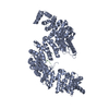
 Movie
Movie Controller


 Open data
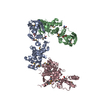
 Basic information
 Components
 Keywords
 Function and homology information
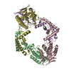
 Streptococcus agalactiae (bacteria)
 X-RAY DIFFRACTION /
  Authors
 Citation
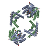
 Structure visualization
 Downloads & links
 Other downloads
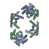
 PDBj
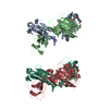
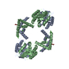
PDBj Assembly
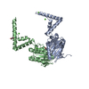

 Mass: 40.078 Da / Num. of mol.: 5 / Source method: obtained synthetically / Formula: Ca
 Mass: 62.068 Da / Num. of mol.: 3 / Source method: obtained synthetically / Formula: C2H6O2
 Mass: 18.015 Da / Num. of mol.: 296 / Source method: isolated from a natural source / Formula: H2O
 Sample preparation
 / Beamline: BM30A / Wavelength: 0.979981 Å
 Processing