 Movie
Movie Controller

[English] 日本語
 Yorodumi
- PDB-3qfi: X-ray crystal structure of transcriptional regulator (EF0465) fro... -
+ Open data

- Basic information
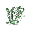
| Entry | Database: PDB / ID: 3qfi | ||||||
|---|---|---|---|---|---|---|---|
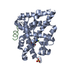
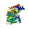
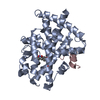
| Title | X-ray crystal structure of transcriptional regulator (EF0465) from Enterococcus faecalis, Northeast Structural Genomics Consortium Target EfR190 | ||||||
 Components Components | Transcriptional regulator | ||||||
 Keywords Keywords | TRANSCRIPTION REGULATOR / Structural Genomics / PSI-Biology / Protein Structure Initiative / Northeast Structural Genomics Consortium / NESG / alpha-beta protein / transcriptional regulator | ||||||
| Function / homology | LCP protein / : / Cell envelope-related transcriptional attenuator domain / LytR_cpsA_psr family / Aminopeptidase / 3-Layer(aba) Sandwich / Alpha Beta / Transcriptional regulator Function and homology information Function and homology information | ||||||
| Biological species |   Enterococcus faecalis (bacteria) Enterococcus faecalis (bacteria) | ||||||
| Method |  X-RAY DIFFRACTION / SYNCHROTRON / MOLECULAR REPLACEMENT / Resolution: 2.71 Å X-RAY DIFFRACTION / SYNCHROTRON / MOLECULAR REPLACEMENT / Resolution: 2.71 Å | ||||||
 Authors Authors | Forouhar, F. / Neely, H. / Seetharaman, J. / Wang, H. / Ciccosanti, C. / Mao, L. / Acton, T.B. / Xiao, R. / Everett, J.K. / Montelione, G.T. ...Forouhar, F. / Neely, H. / Seetharaman, J. / Wang, H. / Ciccosanti, C. / Mao, L. / Acton, T.B. / Xiao, R. / Everett, J.K. / Montelione, G.T. / Tong, L. / Hun, J.F. / Northeast Structural Genomics Consortium (NESG) | ||||||
 Citation Citation | Journal: To be Published Title: X-ray crystal structure of transcriptional regulator (EF0465) from Enterococcus faecalis, Northeast Structural Genomics Consortium Target EfR190 Authors: Forouhar, F. / Neely, H. / Seetharaman, J. / Wang, H. / Ciccosanti, C. / Mao, L. / Acton, T.B. / Xiao, R. / Everett, J.K. / Montelione, G.T. / Tong, L. / Hun, J.F. / Northeast Structural ...Authors: Forouhar, F. / Neely, H. / Seetharaman, J. / Wang, H. / Ciccosanti, C. / Mao, L. / Acton, T.B. / Xiao, R. / Everett, J.K. / Montelione, G.T. / Tong, L. / Hun, J.F. / Northeast Structural Genomics Consortium (NESG) | ||||||
| History |
|
- Structure visualization
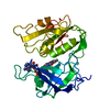
| Structure viewer | Molecule: MolmilJmol/JSmol |
|---|
- Downloads & links
-Download
| PDBx/mmCIF format | 3qfi.cif.gz | 65.7 KB | Display | PDBx/mmCIF format |
|---|---|---|---|---|
| PDB format | pdb3qfi.ent.gz | 47.1 KB | Display | PDB format |
| PDBx/mmJSON format | 3qfi.json.gz | Tree view | PDBx/mmJSON format | |
| Others |  Other downloads Other downloads |
-Validation report
| Arichive directory | https://data.pdbj.org/pub/pdb/validation_reports/qf/3qfiftp://data.pdbj.org/pub/pdb/validation_reports/qf/3qfi | HTTPS FTP |
|---|
-Related structure data
| Related structure data | 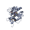 3okzS S: Starting model for refinement |
|---|---|
| Similar structure data | |
| Other databases |


-Links
 PDBj
PDBj- Assembly
| Deposited unit | 
| ||||||||
|---|---|---|---|---|---|---|---|---|---|
| 1 |
| ||||||||
| Unit cell |
|
-Components
| #1: Protein | Mass: 33763.941 Da / Num. of mol.: 1 / Fragment: sequence database residues 42-333 Source method: isolated from a genetically manipulated source Source: (gene. exp.) Enterococcus faecalis (bacteria) / Strain: V583 / Gene: EF0465, EF_0465 / Production host: |
|---|---|
| #2: Water | ChemComp-HOH /  Mass: 18.015 Da / Num. of mol.: 38 / Source method: isolated from a natural source / Formula: H2O Mass: 18.015 Da / Num. of mol.: 38 / Source method: isolated from a natural source / Formula: H2O |
-Experimental details
-Experiment
| Experiment | Method: X-RAY DIFFRACTION / Number of used crystals: 1 |
|---|
- Sample preparation
| Crystal | Density Matthews: 1.9 Å3/Da / Density % sol: 35.2 % |
|---|---|
| Crystal grow | Temperature: 277 K / Method: microbatch under oil method / pH: 7.5 Details: Protein solution: 10 mM Tris (pH 7.5), 100 mM sodium chloride, and 5 mM DTT, Precipitation solution: 0.1M HEPES (pH 7.5), 40% PEG400, and 0.1M calcium acetate, microbatch under oil method, temperature 277K |
-Data collection
| Diffraction | Mean temperature: 100 K |
|---|---|
| Diffraction source | Source: SYNCHROTRON / Site: NSLS  / Beamline: X4C / Wavelength: 0.97877 Å / Beamline: X4C / Wavelength: 0.97877 Å |
| Detector | Type: MAR CCD 165 mm / Detector: CCD / Date: Jun 4, 2010 / Details: mirrors |
| Radiation | Monochromator: Si 111 CHANNEL / Protocol: SINGLE WAVELENGTH / Monochromatic (M) / Laue (L): M / Scattering type: x-ray |
| Radiation wavelength | Wavelength: 0.97877 Å / Relative weight: 1 |
| Reflection | Resolution: 2.7→30 Å / Num. all: 7395 / Num. obs: 6316 / % possible obs: 85.4 % / Observed criterion σ(F): 0.3 / Observed criterion σ(I): 0.3 / Redundancy: 4.3 % / Biso Wilson estimate: 15.9 Å2 / Rmerge(I) obs: 0.074 / Rsym value: 0.078 / Net I/σ(I): 14.88 |
| Reflection shell | Resolution: 2.7→2.8 Å / Redundancy: 2.3 % / Rmerge(I) obs: 0.109 / Mean I/σ(I) obs: 4.3 / Num. unique all: 465 / Rsym value: 0.125 / % possible all: 65.3 |
- Processing
| Software |
| |||||||||||||||||||||||||
|---|---|---|---|---|---|---|---|---|---|---|---|---|---|---|---|---|---|---|---|---|---|---|---|---|---|---|
| Refinement | Method to determine structure: MOLECULAR REPLACEMENT Starting model: 3OKZ Resolution: 2.71→19.87 Å / Rfactor Rfree error: 0.01 / Data cutoff high absF: 406735.4 / Data cutoff low absF: 0 / Isotropic thermal model: RESTRAINED / Cross valid method: THROUGHOUT / σ(F): 2 / σ(I): 2 / Stereochemistry target values: Engh & Huber
| |||||||||||||||||||||||||
| Solvent computation | Solvent model: FLAT MODEL / Bsol: 19.0745 Å2 / ksol: 0.35 e/Å3 | |||||||||||||||||||||||||
| Displacement parameters | Biso mean: 24.5 Å2
| |||||||||||||||||||||||||
| Refine analyze |
| |||||||||||||||||||||||||
| Refinement step | Cycle: LAST / Resolution: 2.71→19.87 Å
| |||||||||||||||||||||||||
| Refine LS restraints |
| |||||||||||||||||||||||||
| Refine LS restraints NCS | NCS model details: NONE | |||||||||||||||||||||||||
| LS refinement shell | Resolution: 2.7→2.8 Å / Rfactor Rfree error: 0.034 / Total num. of bins used: 10
| |||||||||||||||||||||||||
| Xplor file |
|
