 Movie
Movie Controller

[English] 日本語
 Yorodumi
- PDB-3q0x: N-terminal coiled-coil dimer domain of C. reinhardtii SAS-6 homol... -
+ Open data

- Basic information
| Entry | Database: PDB / ID: 3q0x | ||||||
|---|---|---|---|---|---|---|---|
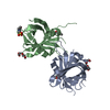
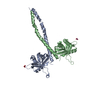
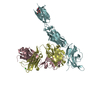
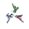
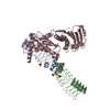
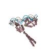
| Title | N-terminal coiled-coil dimer domain of C. reinhardtii SAS-6 homolog Bld12p | ||||||
 Components Components | Centriole protein | ||||||
 Keywords Keywords | STRUCTURAL PROTEIN / centrosome protein / coiled coil mediated dimer | ||||||
| Function / homology |  Function and homology information Function and homology information | ||||||
| Biological species |   Chlamydomonas reinhardtii (plant) Chlamydomonas reinhardtii (plant) | ||||||
| Method |  X-RAY DIFFRACTION / SYNCHROTRON / MOLECULAR REPLACEMENT / Resolution: 3.02 Å X-RAY DIFFRACTION / SYNCHROTRON / MOLECULAR REPLACEMENT / Resolution: 3.02 Å | ||||||
 Authors Authors | Kitagawa, D. / Vakonakis, I. / Olieric, N. / Hilbert, M. / Keller, D. / Olieric, V. / Bortfeld, M. / Erat, M.C. / Flueckiger, I. / Goenczy, P. / Steinmetz, M.O. | ||||||
 Citation Citation | Journal: Cell(Cambridge,Mass.) / Year: 2011 Title: Structural basis of the 9-fold symmetry of centrioles. Authors: Kitagawa, D. / Vakonakis, I. / Olieric, N. / Hilbert, M. / Keller, D. / Olieric, V. / Bortfeld, M. / Erat, M.C. / Fluckiger, I. / Gonczy, P. / Steinmetz, M.O. | ||||||
| History |
|
- Structure visualization
| Structure viewer | Molecule: MolmilJmol/JSmol |
|---|
- Downloads & links
-Download
| PDBx/mmCIF format | 3q0x.cif.gz | 89.6 KB | Display | PDBx/mmCIF format |
|---|---|---|---|---|
| PDB format | pdb3q0x.ent.gz | 67.2 KB | Display | PDB format |
| PDBx/mmJSON format | 3q0x.json.gz | Tree view | PDBx/mmJSON format | |
| Others |  Other downloads Other downloads |
-Validation report
| Arichive directory | https://data.pdbj.org/pub/pdb/validation_reports/q0/3q0xftp://data.pdbj.org/pub/pdb/validation_reports/q0/3q0x | HTTPS FTP |
|---|
-Related structure data


-Links
 PDBj
PDBj- Assembly
| Deposited unit | 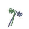
| ||||||||
|---|---|---|---|---|---|---|---|---|---|
| 1 |
| ||||||||
| Unit cell |
|
-Components
| #1: Protein | Mass: 25814.262 Da / Num. of mol.: 2 / Fragment: UNP residues 1-226 / Mutation: F145E Source method: isolated from a genetically manipulated source Details: PSTCM1 pET47B derived / Source: (gene. exp.) Chlamydomonas reinhardtii (plant) / Gene: CrSAS-6, crSAS-6 (Bld12p) / Plasmid: PSTCM1 / Production host:  #2: Water | ChemComp-HOH / |  Mass: 18.015 Da / Num. of mol.: 14 / Source method: isolated from a natural source / Formula: H2O Mass: 18.015 Da / Num. of mol.: 14 / Source method: isolated from a natural source / Formula: H2OHas protein modification | Y | |
|---|
-Experimental details
-Experiment
| Experiment | Method: X-RAY DIFFRACTION / Number of used crystals: 1 |
|---|
- Sample preparation
| Crystal | Density Matthews: 2.73 Å3/Da / Density % sol: 54.91 % |
|---|---|
| Crystal grow | Temperature: 298 K / Method: vapor diffusion / pH: 8.5 Details: 100 mM TrisHCl, pH 8.5, 200 mM MgCl2, 20% PEG8000, 2% benzamidine, vapor diffusion, temperature 298K, VAPOR DIFFUSION |
-Data collection
| Diffraction | Mean temperature: 100 K | |||||||||||||||||||||||||||||||||||||||||||||||||||||||||||||||||||||||||||||
|---|---|---|---|---|---|---|---|---|---|---|---|---|---|---|---|---|---|---|---|---|---|---|---|---|---|---|---|---|---|---|---|---|---|---|---|---|---|---|---|---|---|---|---|---|---|---|---|---|---|---|---|---|---|---|---|---|---|---|---|---|---|---|---|---|---|---|---|---|---|---|---|---|---|---|---|---|---|---|
| Diffraction source | Source: SYNCHROTRON / Site: SLS  / Beamline: X06SA / Wavelength: 1.0015 Å / Beamline: X06SA / Wavelength: 1.0015 Å | |||||||||||||||||||||||||||||||||||||||||||||||||||||||||||||||||||||||||||||
| Detector | Type: MAR225 / Detector: CCD / Date: Oct 10, 2010 | |||||||||||||||||||||||||||||||||||||||||||||||||||||||||||||||||||||||||||||
| Radiation | Protocol: SINGLE WAVELENGTH / Monochromatic (M) / Laue (L): M / Scattering type: x-ray | |||||||||||||||||||||||||||||||||||||||||||||||||||||||||||||||||||||||||||||
| Radiation wavelength | Wavelength: 1.0015 Å / Relative weight: 1 | |||||||||||||||||||||||||||||||||||||||||||||||||||||||||||||||||||||||||||||
| Reflection | Resolution: 3.02→56.29 Å / Num. obs: 10395 / % possible obs: 95.8 % / Observed criterion σ(I): -3 / Redundancy: 11.23 % / Biso Wilson estimate: 59.784 Å2 / Rmerge(I) obs: 0.173 / Net I/σ(I): 10.79 | |||||||||||||||||||||||||||||||||||||||||||||||||||||||||||||||||||||||||||||
| Reflection shell |
|
- Processing
| Software |
| |||||||||||||||||||||||||||||||||||||||||||||||||||||||||
|---|---|---|---|---|---|---|---|---|---|---|---|---|---|---|---|---|---|---|---|---|---|---|---|---|---|---|---|---|---|---|---|---|---|---|---|---|---|---|---|---|---|---|---|---|---|---|---|---|---|---|---|---|---|---|---|---|---|---|
| Refinement | Method to determine structure: MOLECULAR REPLACEMENT / Resolution: 3.02→56.29 Å / Cor.coef. Fo:Fc: 0.912 / Cor.coef. Fo:Fc free: 0.8665 / Occupancy max: 1 / Occupancy min: 1 / Cross valid method: THROUGHOUT / σ(F): 0
| |||||||||||||||||||||||||||||||||||||||||||||||||||||||||
| Displacement parameters | Biso max: 137.35 Å2 / Biso mean: 68.0898 Å2 / Biso min: 33.43 Å2
| |||||||||||||||||||||||||||||||||||||||||||||||||||||||||
| Refine analyze | Luzzati coordinate error obs: 0.583 Å | |||||||||||||||||||||||||||||||||||||||||||||||||||||||||
| Refinement step | Cycle: LAST / Resolution: 3.02→56.29 Å
| |||||||||||||||||||||||||||||||||||||||||||||||||||||||||
| Refine LS restraints |
| |||||||||||||||||||||||||||||||||||||||||||||||||||||||||
| LS refinement shell | Resolution: 3.02→3.38 Å / Total num. of bins used: 5
|
