 Movie
Movie Controller

[English] 日本語
 Yorodumi
- PDB-3pzi: Structure of the hyperthermostable endo-1,4-beta-D-mannanase from... -
+ Open data

- Basic information
| Entry | Database: PDB / ID: 3pzi | ||||||
|---|---|---|---|---|---|---|---|
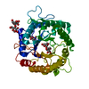
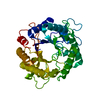
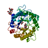
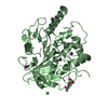
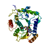
| Title | Structure of the hyperthermostable endo-1,4-beta-D-mannanase from Thermotoga petrophila RKU-1 in complex with beta-D-glucose | ||||||
 Components Components | Mannan endo-1,4-beta-mannosidase. Glycosyl Hydrolase family 5 | ||||||
 Keywords Keywords | HYDROLASE / alpha/beta barrel / glycosyl hydrolase / sugar binding / secreted | ||||||
| Function / homology |  Function and homology information Function and homology informationmannan endo-1,4-beta-mannosidase / mannan endo-1,4-beta-mannosidase activity Similarity search - Function | ||||||
| Biological species |   Thermotoga petrophila (bacteria) Thermotoga petrophila (bacteria) | ||||||
| Method |  X-RAY DIFFRACTION / SYNCHROTRON / MOLECULAR REPLACEMENT / Resolution: 1.55 Å X-RAY DIFFRACTION / SYNCHROTRON / MOLECULAR REPLACEMENT / Resolution: 1.55 Å | ||||||
 Authors Authors | Santos, C.R. / Meza, A.N. / Paiva, J.H. / Silva, J.C. / Ruller, R. / Prade, R.A. / Squina, F.M. / Murakami, M.T. | ||||||
 Citation Citation | Journal: To be Published Title: Structural characterization of a novel hyperthermostable endo-1,4-beta-D-mannanase from Thermotoga petrophila RKU-1 Authors: Santos, C.R. / Meza, A.N. / Paiva, J.H. / Silva, J.C. / Ruller, R. / Prade, R.A. / Squina, F.M. / Murakami, M.T. | ||||||
| History |
|
- Structure visualization
| Structure viewer | Molecule: MolmilJmol/JSmol |
|---|
- Downloads & links
-Download
| PDBx/mmCIF format | 3pzi.cif.gz | 172.9 KB | Display | PDBx/mmCIF format |
|---|---|---|---|---|
| PDB format | pdb3pzi.ent.gz | 136.4 KB | Display | PDB format |
| PDBx/mmJSON format | 3pzi.json.gz | Tree view | PDBx/mmJSON format | |
| Others |  Other downloads Other downloads |
-Validation report
| Summary document | 3pzi_validation.pdf.gz | 448.4 KB | Display | wwPDB validaton report |
|---|---|---|---|---|
| Full document | 3pzi_full_validation.pdf.gz | 452.4 KB | Display | |
| Data in XML | 3pzi_validation.xml.gz | 18.9 KB | Display | |
| Data in CIF | 3pzi_validation.cif.gz | 29.2 KB | Display | |
| Arichive directory | https://data.pdbj.org/pub/pdb/validation_reports/pz/3pziftp://data.pdbj.org/pub/pdb/validation_reports/pz/3pzi | HTTPS FTP |
-Related structure data
| Related structure data |  3pz9C  3pzgC 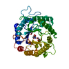 3pzmC  3pznC  3pzoC  3pzqC C: citing same article ( |
|---|---|
| Similar structure data |


-Links
 PDBj
PDBj


- Assembly
| Deposited unit | 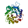
| ||||||||
|---|---|---|---|---|---|---|---|---|---|
| 1 |
| ||||||||
| Unit cell |
|
-Components
| #1: Protein | Mass: 44213.379 Da / Num. of mol.: 1 / Fragment: UNP residues 32-393 Source method: isolated from a genetically manipulated source Source: (gene. exp.) Thermotoga petrophila (bacteria) / Strain: RKU-1 / Gene: Tpet_1542 / Production host: References: UniProt: A5IMX7, mannan endo-1,4-beta-mannosidase |
|---|---|
| #2: Sugar | ChemComp-BGC /   Type: D-saccharide, beta linking / Mass: 180.156 Da / Num. of mol.: 1 Type: D-saccharide, beta linking / Mass: 180.156 Da / Num. of mol.: 1Source method: isolated from a genetically manipulated source Formula: C6H12O6 |
| #3: Water | ChemComp-HOH /  Mass: 18.015 Da / Num. of mol.: 370 / Source method: isolated from a natural source / Formula: H2O Mass: 18.015 Da / Num. of mol.: 370 / Source method: isolated from a natural source / Formula: H2O |
-Experimental details
-Experiment
| Experiment | Method: X-RAY DIFFRACTION / Number of used crystals: 1 |
|---|
- Sample preparation
| Crystal | Density Matthews: 2.4 Å3/Da / Density % sol: 48.7 % |
|---|---|
| Crystal grow | Temperature: 291 K / Method: vapor diffusion, sitting drop / pH: 5.5 Details: 0.8 M phosphate, 0.2 M sodium chloride, pH 5.5, VAPOR DIFFUSION, SITTING DROP, temperature 291K |
-Data collection
| Diffraction | Mean temperature: 100 K |
|---|---|
| Diffraction source | Source: SYNCHROTRON / Site: LNLS  / Beamline: W01B-MX2 / Wavelength: 1.4586 Å / Beamline: W01B-MX2 / Wavelength: 1.4586 Å |
| Detector | Type: MARMOSAIC 225 mm CCD / Detector: CCD / Date: Apr 10, 2010 |
| Radiation | Monochromator: Si(111) double crystal / Protocol: SINGLE WAVELENGTH / Monochromatic (M) / Laue (L): M / Scattering type: x-ray |
| Radiation wavelength | Wavelength: 1.4586 Å / Relative weight: 1 |
| Reflection | Resolution: 1.55→35.4 Å / Num. obs: 62462 / % possible obs: 99.9 % / Observed criterion σ(I): 2 |
| Reflection shell | Resolution: 1.55→1.61 Å / % possible all: 99.1 |
- Processing
| Software |
| ||||||||||||||||||||||||||||||||||||||||||||||||||||||||||||||||||||||||||||||||
|---|---|---|---|---|---|---|---|---|---|---|---|---|---|---|---|---|---|---|---|---|---|---|---|---|---|---|---|---|---|---|---|---|---|---|---|---|---|---|---|---|---|---|---|---|---|---|---|---|---|---|---|---|---|---|---|---|---|---|---|---|---|---|---|---|---|---|---|---|---|---|---|---|---|---|---|---|---|---|---|---|---|
| Refinement | Method to determine structure: MOLECULAR REPLACEMENT / Resolution: 1.55→35.4 Å / Cor.coef. Fo:Fc: 0.974 / Cor.coef. Fo:Fc free: 0.96 / SU B: 2.366 / SU ML: 0.039 / Cross valid method: THROUGHOUT / ESU R Free: 0.068 / Stereochemistry target values: MAXIMUM LIKELIHOOD
| ||||||||||||||||||||||||||||||||||||||||||||||||||||||||||||||||||||||||||||||||
| Solvent computation | Ion probe radii: 0.8 Å / Shrinkage radii: 0.8 Å / VDW probe radii: 1.4 Å / Solvent model: MASK | ||||||||||||||||||||||||||||||||||||||||||||||||||||||||||||||||||||||||||||||||
| Displacement parameters | Biso mean: 17.022 Å2
| ||||||||||||||||||||||||||||||||||||||||||||||||||||||||||||||||||||||||||||||||
| Refinement step | Cycle: LAST / Resolution: 1.55→35.4 Å
| ||||||||||||||||||||||||||||||||||||||||||||||||||||||||||||||||||||||||||||||||
| Refine LS restraints |
| ||||||||||||||||||||||||||||||||||||||||||||||||||||||||||||||||||||||||||||||||
| LS refinement shell | Resolution: 1.55→1.59 Å / Total num. of bins used: 20
|
