 Movie
Movie Controller

[English] 日本語
 Yorodumi
- PDB-3pyd: crystal structure of 4-diphosphocytidyl-2-C-methyl-D-erythritol k... -
+ Open data

- Basic information
| Entry | Database: PDB / ID: 3pyd | ||||||
|---|---|---|---|---|---|---|---|
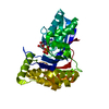
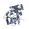
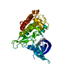
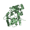
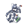
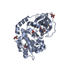
| Title | crystal structure of 4-diphosphocytidyl-2-C-methyl-D-erythritol kinase (IspE) from Mycobacterium tuberculosis | ||||||
 Components Components | 4-diphosphocytidyl-2-C-methyl-D-erythritol kinase | ||||||
 Keywords Keywords | TRANSFERASE / kinase / 4-diphosphocytidyl-2-C-methyl-D-erythritol kinase | ||||||
| Function / homology |  Function and homology information Function and homology information4-(cytidine 5'-diphospho)-2-C-methyl-D-erythritol kinase / 4-(cytidine 5'-diphospho)-2-C-methyl-D-erythritol kinase activity / isopentenyl diphosphate biosynthetic process, methylerythritol 4-phosphate pathway / terpenoid biosynthetic process / ATP binding Similarity search - Function | ||||||
| Biological species |   Mycobacterium tuberculosis (bacteria) Mycobacterium tuberculosis (bacteria) | ||||||
| Method |  X-RAY DIFFRACTION / MOLECULAR REPLACEMENT / Resolution: 2.101 Å X-RAY DIFFRACTION / MOLECULAR REPLACEMENT / Resolution: 2.101 Å | ||||||
 Authors Authors | Shan, S. / Chen, X.H. / Liu, T. / Zhao, H.C. / Rao, Z.H. / Lou, Z.Y. | ||||||
 Citation Citation | Journal: To be Published Title: The Structural Basis for anti-TB Drug Discovery targeting of 4-diphosphocytidyl-2-C-methyl-D-erythritol kinase (IspE) from Mycobacterium tuberculosis Authors: Shan, S. / Chen, X.H. / Liu, T. / Zhao, H.C. / Rao, Z.H. / Lou, Z.Y. | ||||||
| History |
|
- Structure visualization
| Structure viewer | Molecule: MolmilJmol/JSmol |
|---|
- Downloads & links
-Download
| PDBx/mmCIF format | 3pyd.cif.gz | 71.7 KB | Display | PDBx/mmCIF format |
|---|---|---|---|---|
| PDB format | pdb3pyd.ent.gz | 51.9 KB | Display | PDB format |
| PDBx/mmJSON format | 3pyd.json.gz | Tree view | PDBx/mmJSON format | |
| Others |  Other downloads Other downloads |
-Validation report
| Arichive directory | https://data.pdbj.org/pub/pdb/validation_reports/py/3pydftp://data.pdbj.org/pub/pdb/validation_reports/py/3pyd | HTTPS FTP |
|---|
-Related structure data
| Related structure data |  3pyeC  3pyfC 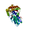 3pygC 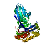 1uekS C: citing same article ( S: Starting model for refinement |
|---|---|
| Similar structure data |

-Links
 PDBj
PDBj- Assembly
| Deposited unit | 
| ||||||||
|---|---|---|---|---|---|---|---|---|---|
| 1 |
| ||||||||
| Unit cell |
|
-Components
| #1: Protein | Mass: 31408.590 Da / Num. of mol.: 1 Source method: isolated from a genetically manipulated source Source: (gene. exp.) Mycobacterium tuberculosis (bacteria) / Gene: ispE, Rv1011, MT1040, MTCI237.28 / Production host: References: UniProt: P65178, UniProt: P9WKG7*PLUS, 4-(cytidine 5'-diphospho)-2-C-methyl-D-erythritol kinase |
|---|---|
| #2: Chemical | ChemComp-GOL /   Mass: 92.094 Da / Num. of mol.: 1 / Source method: obtained synthetically / Formula: C3H8O3 Mass: 92.094 Da / Num. of mol.: 1 / Source method: obtained synthetically / Formula: C3H8O3 |
| #3: Water | ChemComp-HOH /  Mass: 18.015 Da / Num. of mol.: 190 / Source method: isolated from a natural source / Formula: H2O Mass: 18.015 Da / Num. of mol.: 190 / Source method: isolated from a natural source / Formula: H2O |
-Experimental details
-Experiment
| Experiment | Method: X-RAY DIFFRACTION / Number of used crystals: 1 |
|---|
- Sample preparation
| Crystal | Density Matthews: 3.25 Å3/Da / Density % sol: 62.19 % |
|---|---|
| Crystal grow | Temperature: 291 K / Method: vapor diffusion, hanging drop / pH: 7 Details: 0.1M Bis-Tris propane (pH 7.0), 3.2M NaCl, VAPOR DIFFUSION, HANGING DROP, temperature 291K |
-Data collection
| Diffraction | Mean temperature: 100 K |
|---|---|
| Diffraction source | Source: ROTATING ANODE / Type: RIGAKU FR-E SUPERBRIGHT / Wavelength: 1.5418 Å |
| Detector | Type: RIGAKU RAXIS IV++ / Detector: IMAGE PLATE / Date: Jul 8, 2009 |
| Radiation | Protocol: SINGLE WAVELENGTH / Monochromatic (M) / Laue (L): M / Scattering type: x-ray |
| Radiation wavelength | Wavelength: 1.5418 Å / Relative weight: 1 |
| Reflection | Resolution: 2.1→50 Å / Num. all: 24625 / Num. obs: 24569 / % possible obs: 99 % / Observed criterion σ(F): 0 / Observed criterion σ(I): 0 |
- Processing
| Software |
| ||||||||||||||||||||||||||||||||||||||||||||||||||||||||||||
|---|---|---|---|---|---|---|---|---|---|---|---|---|---|---|---|---|---|---|---|---|---|---|---|---|---|---|---|---|---|---|---|---|---|---|---|---|---|---|---|---|---|---|---|---|---|---|---|---|---|---|---|---|---|---|---|---|---|---|---|---|---|
| Refinement | Method to determine structure: MOLECULAR REPLACEMENT Starting model: 1UEK Resolution: 2.101→37.57 Å / SU ML: 0.32 / σ(F): 0.12 / Stereochemistry target values: ML
| ||||||||||||||||||||||||||||||||||||||||||||||||||||||||||||
| Solvent computation | Shrinkage radii: 0.9 Å / VDW probe radii: 1.11 Å / Solvent model: FLAT BULK SOLVENT MODEL / Bsol: 57.848 Å2 / ksol: 0.395 e/Å3 | ||||||||||||||||||||||||||||||||||||||||||||||||||||||||||||
| Displacement parameters |
| ||||||||||||||||||||||||||||||||||||||||||||||||||||||||||||
| Refinement step | Cycle: LAST / Resolution: 2.101→37.57 Å
| ||||||||||||||||||||||||||||||||||||||||||||||||||||||||||||
| Refine LS restraints |
| ||||||||||||||||||||||||||||||||||||||||||||||||||||||||||||
| LS refinement shell | Refine-ID: X-RAY DIFFRACTION / Total num. of bins used: 9
|
