[English] 日本語
 Yorodumi
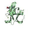
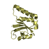
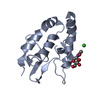
- PDB-3ph0: Crystal structure of the heteromolecular chaperone, AscE-AscG, fr... -
+ Open data
- Basic information
| Entry | Database: PDB / ID: 3ph0 | ||||||
|---|---|---|---|---|---|---|---|
| Title | Crystal structure of the heteromolecular chaperone, AscE-AscG, from the type III secretion system in Aeromonas hydrophila | ||||||
 Components Components |
| ||||||
 Keywords Keywords | CHAPERONE / Type III secretion system / chaperones AscE and AscG | ||||||
| Function / homology |  Function and homology information Function and homology informationType III secretion system YscG / Bacterial type III secretion system chaperone protein / Type III secretion system, secretion protein E / Type III secretion system, cytoplasmic E component of needle / Serine Threonine Protein Phosphatase 5, Tetratricopeptide repeat - #1040 / Immunoglobulin FC, subunit C / Single alpha-helices involved in coiled-coils or other helix-helix interfaces / Serine Threonine Protein Phosphatase 5, Tetratricopeptide repeat / Alpha Horseshoe / Tetratricopeptide-like helical domain superfamily ...Type III secretion system YscG / Bacterial type III secretion system chaperone protein / Type III secretion system, secretion protein E / Type III secretion system, cytoplasmic E component of needle / Serine Threonine Protein Phosphatase 5, Tetratricopeptide repeat - #1040 / Immunoglobulin FC, subunit C / Single alpha-helices involved in coiled-coils or other helix-helix interfaces / Serine Threonine Protein Phosphatase 5, Tetratricopeptide repeat / Alpha Horseshoe / Tetratricopeptide-like helical domain superfamily / Up-down Bundle / Mainly Alpha Similarity search - Domain/homology | ||||||
| Biological species |  Aeromonas hydrophila (bacteria) Aeromonas hydrophila (bacteria) | ||||||
| Method |  X-RAY DIFFRACTION / X-RAY DIFFRACTION /  SYNCHROTRON / SYNCHROTRON /  MAD / Resolution: 2.4 Å MAD / Resolution: 2.4 Å | ||||||
 Authors Authors | Chatterjee, C. / Kumar, S. / Chakraborty, S. / Tan, Y.W. / Leung, K.Y. / Sivaraman, J. / Mok, Y.K. | ||||||
 Citation Citation |  Journal: Plos One / Year: 2011 Journal: Plos One / Year: 2011Title: Crystal structure of the heteromolecular chaperone, AscE-AscG, from the type III secretion system in Aeromonas hydrophila Authors: Chatterjee, C. / Kumar, S. / Chakraborty, S. / Tan, Y.W. / Leung, K.Y. / Sivaraman, J. / Mok, Y.K. | ||||||
| History |
|
- Structure visualization
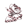
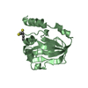
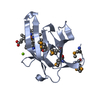
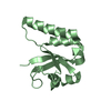
| Structure viewer | Molecule:  Molmil Molmil Jmol/JSmol Jmol/JSmol |
|---|
- Downloads & links
- Download
| PDBx/mmCIF format |  3ph0.cif.gz 3ph0.cif.gz | 59.8 KB | Display |  PDBx/mmCIF format PDBx/mmCIF format |
|---|---|---|---|---|
| PDB format |  pdb3ph0.ent.gz pdb3ph0.ent.gz | 44.5 KB | Display |  PDB format PDB format |
| PDBx/mmJSON format |  3ph0.json.gz 3ph0.json.gz | Tree view |  PDBx/mmJSON format PDBx/mmJSON format | |
| Others |  Other downloads Other downloads |
-Validation report
| Summary document |  3ph0_validation.pdf.gz 3ph0_validation.pdf.gz | 449.4 KB | Display |  wwPDB validaton report wwPDB validaton report |
|---|---|---|---|---|
| Full document |  3ph0_full_validation.pdf.gz 3ph0_full_validation.pdf.gz | 459.3 KB | Display | |
| Data in XML |  3ph0_validation.xml.gz 3ph0_validation.xml.gz | 15.1 KB | Display | |
| Data in CIF |  3ph0_validation.cif.gz 3ph0_validation.cif.gz | 20.9 KB | Display | |
| Arichive directory |  https://data.pdbj.org/pub/pdb/validation_reports/ph/3ph0 https://data.pdbj.org/pub/pdb/validation_reports/ph/3ph0 ftp://data.pdbj.org/pub/pdb/validation_reports/ph/3ph0 ftp://data.pdbj.org/pub/pdb/validation_reports/ph/3ph0 | HTTPS FTP |
-Related structure data
| Similar structure data |
|---|
- Links
Links
- Assembly
| Deposited unit | 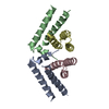
| ||||||||
|---|---|---|---|---|---|---|---|---|---|
| 1 | 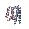
| ||||||||
| 2 | 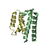
| ||||||||
| Unit cell |
|
- Components
| #1: Protein | Mass: 7572.576 Da / Num. of mol.: 2 Source method: isolated from a genetically manipulated source Source: (gene. exp.)  Aeromonas hydrophila (bacteria) / Production host: Aeromonas hydrophila (bacteria) / Production host:  #2: Protein | Mass: 6760.776 Da / Num. of mol.: 2 / Fragment: residues 1-61 Source method: isolated from a genetically manipulated source Source: (gene. exp.)  Aeromonas hydrophila (bacteria) / Production host: Aeromonas hydrophila (bacteria) / Production host:  #3: Water | ChemComp-HOH / | |
|---|
-Experimental details
-Experiment
| Experiment | Method:  X-RAY DIFFRACTION / Number of used crystals: 1 X-RAY DIFFRACTION / Number of used crystals: 1 |
|---|
- Sample preparation
| Crystal | Density Matthews: 2.35 Å3/Da / Density % sol: 47.62 % |
|---|---|
| Crystal grow | Temperature: 298 K / Method: vapor diffusion, hanging drop / pH: 7.5 Details: 10mM Tris(pH 7.4), 5mM DTT, VAPOR DIFFUSION, HANGING DROP, temperature 298K |
-Data collection
| Diffraction | Mean temperature: 100 K | |||||||||||||||
|---|---|---|---|---|---|---|---|---|---|---|---|---|---|---|---|---|
| Diffraction source | Source:  SYNCHROTRON / Site: SYNCHROTRON / Site:  NSRRC NSRRC  / Beamline: BL13B1 / Wavelength: 0.9792, 0.9794, 0.9640, 1.542 / Beamline: BL13B1 / Wavelength: 0.9792, 0.9794, 0.9640, 1.542 | |||||||||||||||
| Detector | Type: ADSC QUANTUM 315 / Detector: CCD / Date: Dec 1, 2009 | |||||||||||||||
| Radiation | Protocol: MAD / Monochromatic (M) / Laue (L): M / Scattering type: x-ray | |||||||||||||||
| Radiation wavelength |
| |||||||||||||||
| Reflection | Resolution: 2.4→50 Å / Num. obs: 16865 / % possible obs: 99.8 % / Rsym value: 0.089 | |||||||||||||||
| Reflection shell | Highest resolution: 2.4 Å / Rsym value: 0.089 / % possible all: 99.8 |
-Phasing
| Phasing | Method:  MAD MAD |
|---|
- Processing
| Software |
| ||||||||||||||||||||||||||||||||
|---|---|---|---|---|---|---|---|---|---|---|---|---|---|---|---|---|---|---|---|---|---|---|---|---|---|---|---|---|---|---|---|---|---|
| Refinement | Method to determine structure:  MAD / Resolution: 2.4→20 Å / Occupancy max: 1 / Occupancy min: 1 MAD / Resolution: 2.4→20 Å / Occupancy max: 1 / Occupancy min: 1
| ||||||||||||||||||||||||||||||||
| Solvent computation | Bsol: 51.8285 Å2 | ||||||||||||||||||||||||||||||||
| Displacement parameters | Biso max: 105.82 Å2 / Biso mean: 44.3555 Å2 / Biso min: 20.74 Å2
| ||||||||||||||||||||||||||||||||
| Refinement step | Cycle: LAST / Resolution: 2.4→20 Å
| ||||||||||||||||||||||||||||||||
| Refine LS restraints |
| ||||||||||||||||||||||||||||||||
| Xplor file |
|
 Movie
Movie Controller




 PDBj
PDBj

