 Movie
Movie Controller

[English] 日本語
 Yorodumi
- PDB-3pfz: Crystal structure of Cel7A from Talaromyces emersonii in complex ... -
+ Open data

- Basic information
| Entry | Database: PDB / ID: 3pfz | ||||||||||||
|---|---|---|---|---|---|---|---|---|---|---|---|---|---|
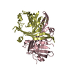
| Title | Crystal structure of Cel7A from Talaromyces emersonii in complex with cellotetraose | ||||||||||||
 Components Components | Cellobiohydrolase 1 catalytic domain | ||||||||||||
 Keywords Keywords | HYDROLASE / cellobiohydrolase / cellulose | ||||||||||||
| Function / homology |  Function and homology information Function and homology informationcellulose 1,4-beta-cellobiosidase activity / Hydrolases; Glycosylases; Glycosidases, i.e. enzymes that hydrolyse O- and S-glycosyl compounds / cellulose catabolic process Similarity search - Function | ||||||||||||
| Biological species |  Talaromyces emersonii (fungus) Talaromyces emersonii (fungus) | ||||||||||||
| Method |  X-RAY DIFFRACTION / SYNCHROTRON / MOLECULAR REPLACEMENT / Resolution: 1.0993 Å X-RAY DIFFRACTION / SYNCHROTRON / MOLECULAR REPLACEMENT / Resolution: 1.0993 Å | ||||||||||||
 Authors Authors | Qin, L. / Pereira, J.H. / McAndrew, R.P. / Simmons, B.A. / Sapra, R. / Adams, P.D. / Sale, K.L. | ||||||||||||
 Citation Citation | Journal: TO BE PUBLISHED Title: Crystal structure of Cel7A from Talaromyces emersonii in complex with cellotetraose Authors: Qin, L. / Pereira, J.H. / McAndrew, R.P. / Simmons, B.A. / Sapra, R. / Adams, P.D. / Sale, K.L. | ||||||||||||
| History |
|
- Structure visualization
| Structure viewer | Molecule: MolmilJmol/JSmol |
|---|
- Downloads & links
-Download
| PDBx/mmCIF format | 3pfz.cif.gz | 285.9 KB | Display | PDBx/mmCIF format |
|---|---|---|---|---|
| PDB format | pdb3pfz.ent.gz | 234.7 KB | Display | PDB format |
| PDBx/mmJSON format | 3pfz.json.gz | Tree view | PDBx/mmJSON format | |
| Others |  Other downloads Other downloads |
-Validation report
| Arichive directory | https://data.pdbj.org/pub/pdb/validation_reports/pf/3pfzftp://data.pdbj.org/pub/pdb/validation_reports/pf/3pfz | HTTPS FTP |
|---|
-Related structure data
| Related structure data |  3pfjS S: Starting model for refinement |
|---|---|
| Similar structure data |


-Links
 PDBj
PDBj- Assembly
| Deposited unit | 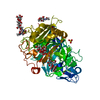
| ||||||||
|---|---|---|---|---|---|---|---|---|---|
| 1 |
| ||||||||
| Unit cell |
|
-Components
-Protein , 1 types, 1 molecules A
| #1: Protein | Mass: 46849.668 Da / Num. of mol.: 1 / Fragment: UNP Residues 19-455 Source method: isolated from a genetically manipulated source Source: (gene. exp.) Talaromyces emersonii (fungus) / Gene: cbh1, cbh1A / Production host: References: UniProt: Q8TFL9, cellulose 1,4-beta-cellobiosidase (non-reducing end) |
|---|
-Sugars , 4 types, 4 molecules 
| #2: Polysaccharide | alpha-D-mannopyranose-(1-3)-beta-D-mannopyranose-(1-4)-2-acetamido-2-deoxy-beta-D-glucopyranose-(1- ...alpha-D-mannopyranose-(1-3)-beta-D-mannopyranose-(1-4)-2-acetamido-2-deoxy-beta-D-glucopyranose-(1-4)-2-acetamido-2-deoxy-beta-D-glucopyranose Source method: isolated from a genetically manipulated source |
|---|---|
| #3: Polysaccharide | beta-D-glucopyranose-(1-4)-beta-D-glucopyranose-(1-4)-beta-D-glucopyranose-(1-4)-beta-D-glucopyranose / beta-cellotetraose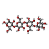  Source method: isolated from a genetically manipulated source Details: oligosaccharide / References: beta-cellotetraose |
| #4: Polysaccharide | beta-D-glucopyranose-(1-4)-beta-D-glucopyranose / beta-cellobiose  Source method: isolated from a genetically manipulated source Details: oligosaccharide / References: beta-cellobiose |
| #5: Sugar | ChemComp-NAG /  Type: D-saccharide, beta linking / Mass: 221.208 Da / Num. of mol.: 1 Type: D-saccharide, beta linking / Mass: 221.208 Da / Num. of mol.: 1Source method: isolated from a genetically manipulated source Formula: C8H15NO6 |
-Non-polymers , 2 types, 868 molecules 

| #6: Chemical |  Mass: 96.063 Da / Num. of mol.: 2 / Source method: obtained synthetically / Formula: SO4 Mass: 96.063 Da / Num. of mol.: 2 / Source method: obtained synthetically / Formula: SO4#7: Water | ChemComp-HOH / | Mass: 18.015 Da / Num. of mol.: 866 / Source method: isolated from a natural source / Formula: H2O |
|---|
-Details
| Has protein modification | Y |
|---|
-Experimental details
-Experiment
| Experiment | Method: X-RAY DIFFRACTION / Number of used crystals: 1 |
|---|
- Sample preparation
| Crystal | Density Matthews: 2.53 Å3/Da / Density % sol: 51.41 % |
|---|---|
| Crystal grow | Temperature: 277 K / Method: vapor diffusion, hanging drop / pH: 9.5 Details: 23 mg/ml Cel7A, 1.8-2.0 M ammonium sulfate, 0.1 M CAPSO pH 9.5, 10 mM cellotetraose, VAPOR DIFFUSION, HANGING DROP, temperature 277K |
-Data collection
| Diffraction | Mean temperature: 100 K |
|---|---|
| Diffraction source | Source: SYNCHROTRON / Site: ALS  / Beamline: 8.2.1 / Wavelength: 0.97 Å / Beamline: 8.2.1 / Wavelength: 0.97 Å |
| Detector | Type: ADSC QUANTUM 315 / Detector: CCD / Date: Oct 30, 2009 |
| Radiation | Monochromator: double crystal, Si(111) / Protocol: SINGLE WAVELENGTH / Monochromatic (M) / Laue (L): M / Scattering type: x-ray |
| Radiation wavelength | Wavelength: 0.97 Å / Relative weight: 1 |
| Reflection | Resolution: 1.0993→28.8 Å / Num. all: 194761 / Num. obs: 185802 / % possible obs: 95.4 % / Observed criterion σ(F): 0 / Observed criterion σ(I): 0 / Redundancy: 7.6 % / Rmerge(I) obs: 0.064 / Net I/σ(I): 25.2 |
| Reflection shell | Resolution: 1.0993→1.14 Å / Redundancy: 5.3 % / Rmerge(I) obs: 0.247 / Mean I/σ(I) obs: 6.3 / Num. unique all: 17259 / % possible all: 89.6 |
- Processing
| Software |
| |||||||||||||||||||||||||||||||||||||||||||||||||||||||||||||||||||||||||||||||||||||||||||||||||||||||||
|---|---|---|---|---|---|---|---|---|---|---|---|---|---|---|---|---|---|---|---|---|---|---|---|---|---|---|---|---|---|---|---|---|---|---|---|---|---|---|---|---|---|---|---|---|---|---|---|---|---|---|---|---|---|---|---|---|---|---|---|---|---|---|---|---|---|---|---|---|---|---|---|---|---|---|---|---|---|---|---|---|---|---|---|---|---|---|---|---|---|---|---|---|---|---|---|---|---|---|---|---|---|---|---|---|---|---|
| Refinement | Method to determine structure: MOLECULAR REPLACEMENT Starting model: PDB ENTRY 3PFJ Resolution: 1.0993→28.78 Å / SU ML: 0.13 / σ(F): 0 / Phase error: 9.68 / Stereochemistry target values: Engh & Huber
| |||||||||||||||||||||||||||||||||||||||||||||||||||||||||||||||||||||||||||||||||||||||||||||||||||||||||
| Solvent computation | Shrinkage radii: 0.9 Å / VDW probe radii: 1.11 Å / Solvent model: FLAT BULK SOLVENT MODEL / Bsol: 39.756 Å2 / ksol: 0.4 e/Å3 | |||||||||||||||||||||||||||||||||||||||||||||||||||||||||||||||||||||||||||||||||||||||||||||||||||||||||
| Displacement parameters |
| |||||||||||||||||||||||||||||||||||||||||||||||||||||||||||||||||||||||||||||||||||||||||||||||||||||||||
| Refinement step | Cycle: LAST / Resolution: 1.0993→28.78 Å
| |||||||||||||||||||||||||||||||||||||||||||||||||||||||||||||||||||||||||||||||||||||||||||||||||||||||||
| Refine LS restraints |
| |||||||||||||||||||||||||||||||||||||||||||||||||||||||||||||||||||||||||||||||||||||||||||||||||||||||||
| LS refinement shell |
|
