 Movie
Movie Controller


+ Open data

- Basic information
| Entry | Database: PDB / ID: 3opb | ||||||
|---|---|---|---|---|---|---|---|
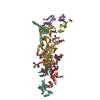
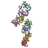
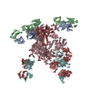
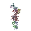
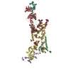
| Title | Crystal structure of She4p | ||||||
 Components Components | SWI5-dependent HO expression protein 4 | ||||||
 Keywords Keywords | PROTEIN BINDING / HEAT and ARM fold / Myosin folding and function / myosin binding protein | ||||||
| Function / homology |  Function and homology information Function and homology informationmating type switching / intracellular mRNA localization / myosin binding / Hsp90 protein binding / actin cytoskeleton organization / identical protein binding / cytoplasm Similarity search - Function | ||||||
| Biological species |  | ||||||
| Method |  X-RAY DIFFRACTION / SYNCHROTRON / MAD / Resolution: 2.9 Å X-RAY DIFFRACTION / SYNCHROTRON / MAD / Resolution: 2.9 Å | ||||||
 Authors Authors | Shi, H. / Blobel, G. | ||||||
 Citation Citation | Journal: Proc.Natl.Acad.Sci.USA / Year: 2010 Title: UNC-45/CRO1/She4p (UCS) protein forms elongated dimer and joins two myosin heads near their actin binding region. Authors: Shi, H. / Blobel, G. | ||||||
| History |
|
- Structure visualization
| Structure viewer | Molecule: MolmilJmol/JSmol |
|---|
- Downloads & links
-Download
| PDBx/mmCIF format | 3opb.cif.gz | 301.9 KB | Display | PDBx/mmCIF format |
|---|---|---|---|---|
| PDB format | pdb3opb.ent.gz | 242.8 KB | Display | PDB format |
| PDBx/mmJSON format | 3opb.json.gz | Tree view | PDBx/mmJSON format | |
| Others |  Other downloads Other downloads |
-Validation report
| Arichive directory | https://data.pdbj.org/pub/pdb/validation_reports/op/3opbftp://data.pdbj.org/pub/pdb/validation_reports/op/3opb | HTTPS FTP |
|---|
-Related structure data
| Similar structure data |
|---|


-Links
 PDBj
PDBj- Assembly
| Deposited unit | 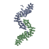
| ||||||||
|---|---|---|---|---|---|---|---|---|---|
| 1 |
| ||||||||
| Unit cell |
|
-Components
| #1: Protein | Mass: 87880.984 Da / Num. of mol.: 2 / Mutation: C4S Source method: isolated from a genetically manipulated source Source: (gene. exp.) Gene: SHE4, YOR035C, OR26.26 / Production host:  #2: Water | ChemComp-HOH / |  Mass: 18.015 Da / Num. of mol.: 141 / Source method: isolated from a natural source / Formula: H2O Mass: 18.015 Da / Num. of mol.: 141 / Source method: isolated from a natural source / Formula: H2OCompound details | THE UNK SEQUENCE CORRESPOND | Sequence details | RESIDUES RANGE 422-440 HAVE POOR DEFINED ELECTRON DENSITY. ONLY 5 RESIDUES CORRESPONDING TO REGION ...RESIDUES RANGE 422-440 HAVE POOR DEFINED ELECTRON DENSITY. ONLY 5 RESIDUES CORRESPOND | |
|---|
-Experimental details
-Experiment
| Experiment | Method: X-RAY DIFFRACTION / Number of used crystals: 1 |
|---|
- Sample preparation
| Crystal | Density Matthews: 2.84 Å3/Da / Density % sol: 56.69 % |
|---|---|
| Crystal grow | Temperature: 285 K / Method: evaporation / pH: 6.5 Details: 200 mM Na citrate, 20% (w/v) PEG 3350, 10 mM DTT, pH 6.5, EVAPORATION, temperature 285K |
-Data collection
| Diffraction |
| ||||||||||||
|---|---|---|---|---|---|---|---|---|---|---|---|---|---|
| Diffraction source |
| ||||||||||||
| Detector |
| ||||||||||||
| Radiation | Monochromator: Si 111 / Protocol: MAD / Monochromatic (M) / Laue (L): M / Scattering type: x-ray | ||||||||||||
| Radiation wavelength | Relative weight: 1 | ||||||||||||
| Reflection | Resolution: 2.9→45 Å / Num. all: 47498 / Num. obs: 45409 / % possible obs: 95.6 % / Observed criterion σ(F): 0 / Observed criterion σ(I): 0 / Rmerge(I) obs: 0.063 | ||||||||||||
| Reflection shell | Resolution: 2.9→3 Å / Redundancy: 6.4 % / Rmerge(I) obs: 0.79 |

- Processing
| Software |
| ||||||||||||||||||||
|---|---|---|---|---|---|---|---|---|---|---|---|---|---|---|---|---|---|---|---|---|---|
| Refinement | Method to determine structure: MAD / Resolution: 2.9→43 Å / σ(F): 0 / Stereochemistry target values: Engh & Huber
| ||||||||||||||||||||
| Refinement step | Cycle: LAST / Resolution: 2.9→43 Å
| ||||||||||||||||||||
| Refine LS restraints |
|
