Mass: 18.015 Da / Num. of mol.: 384 / Source method: isolated from a natural source / Formula: H2O
-
Details
Has protein modification
Y
Sequence details
THIS CONSTRUCT WAS EXPRESSED WITH A PURIFICATION TAG MGSDKIHHHHHHENLYFQG. THE TAG WAS REMOVED WITH ...THIS CONSTRUCT WAS EXPRESSED WITH A PURIFICATION TAG MGSDKIHHHHHHENLYFQG. THE TAG WAS REMOVED WITH TEV PROTEASE LEAVING ONLY A GLYCINE (0) FOLLOWED BY THE TARGET SEQUENCE.
-
Experimental details
-
Experiment
Experiment
Method: X-RAY DIFFRACTION / Number of used crystals: 1
-
Sample preparation
Crystal
Density Matthews: 2.42 Å3/Da / Density % sol: 49.21 %
Crystal grow
Temperature: 293 K / Method: vapor diffusion, sitting drop / pH: 4.6 Details: 1.60M ammonium sulfate, 20.00% Glycerol, 0.1M sodium acetate pH 4.6, NANODROP, VAPOR DIFFUSION, SITTING DROP, temperature 293K
Type: MARMOSAIC 325 mm CCD / Detector: CCD / Date: Jun 10, 2010 / Details: Flat mirror (vertical focusing)
Radiation
Monochromator: Single crystal Si(111) bent monochromator (horizontal focusing) Protocol: MAD / Monochromatic (M) / Laue (L): M / Scattering type: x-ray
Radiation wavelength
ID
Wavelength (Å)
Relative weight
1
0.91837
1
2
0.9792
1
3
0.97889
1
Reflection
Resolution: 1.9→29.197 Å / Num. obs: 52053 / % possible obs: 91.7 % / Observed criterion σ(I): -3 / Biso Wilson estimate: 24.96 Å2 / Rmerge(I) obs: 0.04 / Net I/σ(I): 11.76
Reflection shell
Resolution (Å)
Rmerge(I) obs
Mean I/σ(I) obs
Num. measured obs
Num. unique obs
Diffraction-ID
% possible all
1.9-1.97
0.354
1.8
11111
9387
1
87.9
1.97-2.05
0.238
2.6
11207
9509
1
89.9
2.05-2.14
0.177
3.4
10674
9100
1
90.2
2.14-2.25
0.13
4.6
10870
9270
1
90.9
2.25-2.39
0.095
6
11107
9517
1
91.3
2.39-2.58
0.074
7.7
11529
9942
1
92.1
2.58-2.84
0.051
10.7
11183
9679
1
93.1
2.84-3.25
0.032
16.5
11201
9754
1
93.6
3.25-4.08
0.019
27.5
11100
9713
1
94.1
4.08-29.197
0.014
34.8
11368
9995
1
94.3
-
Phasing
Phasing
Method: MAD
-
Processing
Software
Name
Version
Classification
NB
SHELX
phasing
REFMAC
5.5.0110
refinement
XSCALE
datascaling
PDB_EXTRACT
3.1
dataextraction
XDS
datareduction
SHELXD
phasing
autoSHARP
phasing
Refinement
Method to determine structure: MAD / Resolution: 1.9→29.197 Å / Cor.coef. Fo:Fc: 0.969 / Cor.coef. Fo:Fc free: 0.951 / Occupancy max: 1 / Occupancy min: 0.3 / SU B: 5.576 / SU ML: 0.087 / Cross valid method: THROUGHOUT / σ(F): 0 / ESU R: 0.143 / ESU R Free: 0.129 Stereochemistry target values: MAXIMUM LIKELIHOOD WITH PHASES Details: 1. HYDROGENS HAVE BEEN ADDED IN THE RIDING POSITIONS. 2. ATOM RECORD CONTAINS SUM OF TLS AND RESIDUAL B FACTORS. 3. ANISOU RECORD CONTAINS SUM OF TLS AND RESIDUAL U FACTORS. 4. WATERS WERE ...Details: 1. HYDROGENS HAVE BEEN ADDED IN THE RIDING POSITIONS. 2. ATOM RECORD CONTAINS SUM OF TLS AND RESIDUAL B FACTORS. 3. ANISOU RECORD CONTAINS SUM OF TLS AND RESIDUAL U FACTORS. 4. WATERS WERE EXCLUDED FROM AUTOMATIC TLS ASSIGNMENT. 5. A MET-INHIBITION PROTOCOL WAS USED FOR SELENOMETHIONINE INCORPORATION DURING PROTEIN EXPRESSION. THE OCCUPANCY OF THE SE ATOMS IN THE MSE RESIDUES WAS REDUCED TO 0.75 FOR THE REDUCED SCATTERING POWER DUE TO PARTIAL S-MET INCORPORATION. 6. ACETATE ION (ACT), GLYCEROL (GOL), AND SULFATE ION (SO4) ARE MODELED FROM THE CRYSTALLIZATION SOLUTION. 7. LIGAND MOLECULES FLAVIN-ADENINE DINUCLEOTIDE (FAD) AND FLAVIN MONONUCLEOTIDE (FMN) ARE MODELED BASED ON DENSITY. 8. AN UNKNOWN LIGAND (UNL) HAS BEEN MODELED NEAR FMN IN CHAINS A AND B. THE UNL RESEMBLES ADENINE MOIETY OF FAD AND OCCUPIES NCS RELATED EQUIVALENT POSITION OF THE SAME IN CHAIN C.
Rfactor
Num. reflection
% reflection
Selection details
Rfree
0.1937
2638
5.1 %
RANDOM
Rwork
0.1574
-
-
-
obs
0.1592
52053
98.09 %
-
Solvent computation
Ion probe radii: 0.8 Å / Shrinkage radii: 0.8 Å / VDW probe radii: 1.4 Å / Solvent model: BABINET MODEL WITH MASK
In the structure databanks used in Yorodumi, some data are registered as the other names, "COVID-19 virus" and "2019-nCoV". Here are the details of the virus and the list of structure data.
Jan 31, 2019. EMDB accession codes are about to change! (news from PDBe EMDB page)
EMDB accession codes are about to change! (news from PDBe EMDB page)
The allocation of 4 digits for EMDB accession codes will soon come to an end. Whilst these codes will remain in use, new EMDB accession codes will include an additional digit and will expand incrementally as the available range of codes is exhausted. The current 4-digit format prefixed with “EMD-” (i.e. EMD-XXXX) will advance to a 5-digit format (i.e. EMD-XXXXX), and so on. It is currently estimated that the 4-digit codes will be depleted around Spring 2019, at which point the 5-digit format will come into force.
The EM Navigator/Yorodumi systems omit the EMD- prefix.
Related info.:Q: What is EMD? / ID/Accession-code notation in Yorodumi/EM Navigator
Yorodumi is a browser for structure data from EMDB, PDB, SASBDB, etc.
This page is also the successor to EM Navigator detail page, and also detail information page/front-end page for Omokage search.
The word "yorodu" (or yorozu) is an old Japanese word meaning "ten thousand". "mi" (miru) is to see.
Related info.:EMDB / PDB / SASBDB / Comparison of 3 databanks / Yorodumi Search / Aug 31, 2016. New EM Navigator & Yorodumi / Yorodumi Papers / Jmol/JSmol / Function and homology information / Changes in new EM Navigator and Yorodumi
 Movie
Movie Controller

 Yorodumi
 Open data
 Basic information
 Components
 Keywords
 Function and homology information
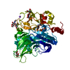
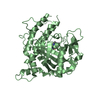
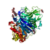
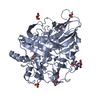
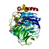
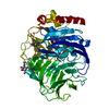
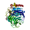
 Idiomarina loihiensis (bacteria)
 X-RAY DIFFRACTION /
  Authors
 Citation
 Structure visualization
 Downloads & links
 Other downloads
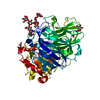


 PDBj
PDBj

 Assembly
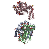


 Mass: 456.344 Da / Num. of mol.: 2 / Source method: obtained synthetically / Formula: C17H21N4O9P
 Mass: 96.063 Da / Num. of mol.: 14 / Source method: obtained synthetically / Formula: SO4
 Mass: 92.094 Da / Num. of mol.: 16 / Source method: obtained synthetically / Formula: C3H8O3
 Mass: 59.044 Da / Num. of mol.: 1 / Source method: obtained synthetically / Formula: C2H3O2
 Mass: 785.550 Da / Num. of mol.: 1 / Source method: obtained synthetically / Formula: C27H33N9O15P2 / Comment: FAD*YM
 Sample preparation
 / Beamline: BL11-1 / Wavelength: 0.91837,0.97920,0.97889
 Processing