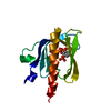
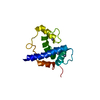
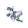
- PDB-3oan: Crystal structure of the Ran Binding Domain From The Nuclear Comp... -
+
Open data
ID or keywords:
Loading...
-
Basic information
Entry
Database: PDB / ID: 3oan
Title
Crystal structure of the Ran Binding Domain From The Nuclear Complex Component Nup2 From Ashbya Gossypii
Components
ABR034Wp
Keywords
TRANSPORT PROTEIN / STRUCTURAL GENOMICS / PROTEIN STRUCTURE INITIATIVE / NEW YORK STRUCTURAL GENOMIX RESEARCH CONSORTIUM / NYSGXRC / PSI-2 / New York SGX Research Center for Structural Genomics
Function / homology
Function and homology information
mRNA export from nucleus in response to heat stress / protein localization to nuclear inner membrane / nuclear pore cytoplasmic filaments / post-transcriptional tethering of RNA polymerase II gene DNA at nuclear periphery / nuclear pore nuclear basket / importin-alpha family protein binding / NLS-dependent protein nuclear import complex / structural constituent of nuclear pore / silent mating-type cassette heterochromatin formation / NLS-bearing protein import into nucleus ...mRNA export from nucleus in response to heat stress / protein localization to nuclear inner membrane / nuclear pore cytoplasmic filaments / post-transcriptional tethering of RNA polymerase II gene DNA at nuclear periphery / nuclear pore nuclear basket / importin-alpha family protein binding / NLS-dependent protein nuclear import complex / structural constituent of nuclear pore / silent mating-type cassette heterochromatin formation / NLS-bearing protein import into nucleus / poly(A)+ mRNA export from nucleus / subtelomeric heterochromatin formation / protein export from nucleus / small GTPase binding / chromosome, telomeric region / cytoplasm Similarity search - Function
Protocol: SINGLE WAVELENGTH / Scattering type: x-ray
Radiation wavelength
Wavelength: 0.9791 Å / Relative weight: 1
Reflection
Resolution: 2.2→50 Å / Num. obs: 15579 / % possible obs: 99.9 % / Redundancy: 7.5 % / Rmerge(I) obs: 0.055 / Net I/σ(I): 12.5
Reflection shell
Resolution (Å)
Redundancy (%)
Rmerge(I) obs
% possible all
2.2-2.24
6.3
0.83
99.4
2.24-2.28
6.8
0.738
99.8
2.28-2.32
7.2
0.623
100
2.32-2.37
7.4
0.518
100
2.37-2.42
7.6
0.457
100
2.42-2.48
7.6
0.365
100
2.48-2.54
7.7
0.319
100
2.54-2.61
7.6
0.266
100
2.61-2.69
7.6
0.231
100
2.69-2.77
7.6
0.207
100
2.77-2.87
7.6
0.146
100
2.87-2.99
7.6
0.124
100
2.99-3.12
7.6
0.088
100
3.12-3.29
7.7
0.067
100
3.29-3.49
7.6
0.054
100
3.49-3.76
7.6
0.049
100
3.76-4.14
7.6
0.043
100
4.14-4.74
7.5
0.036
100
4.74-5.97
7.3
0.03
100
5.97-50
7.6
0.026
98.9
-
Phasing
Phasing
Method: SAD
-
Processing
Software
Name
Version
Classification
NB
SCALEPACK
datascaling
REFMAC
refinement
PDB_EXTRACT
3.1
dataextraction
CBASS
datacollection
HKL-2000
datareduction
PHENIX
phasing
Refinement
Method to determine structure: SAD / Resolution: 2.3→19.98 Å / Cor.coef. Fo:Fc: 0.95 / Cor.coef. Fo:Fc free: 0.896 / Occupancy max: 1 / Occupancy min: 0.5 / SU B: 15.354 / SU ML: 0.171 / SU R Cruickshank DPI: 0.2704 / Cross valid method: THROUGHOUT / ESU R: 0.266 / ESU R Free: 0.248 / Stereochemistry target values: MAXIMUM LIKELIHOOD / Details: HYDROGENS HAVE BEEN ADDED IN THE RIDING POSITIONS
Rfactor
Num. reflection
% reflection
Selection details
Rfree
0.28869
354
4.7 %
RANDOM
Rwork
0.20934
-
-
-
obs
0.21302
7153
99.96 %
-
Solvent computation
Ion probe radii: 0.8 Å / Shrinkage radii: 0.8 Å / VDW probe radii: 1.4 Å / Solvent model: BABINET MODEL WITH MASK
Displacement parameters
Biso mean: 53.948 Å2
Baniso -1
Baniso -2
Baniso -3
1-
0.21 Å2
0 Å2
0 Å2
2-
-
0.21 Å2
0 Å2
3-
-
-
-0.42 Å2
Refinement step
Cycle: LAST / Resolution: 2.3→19.98 Å
Protein
Nucleic acid
Ligand
Solvent
Total
Num. atoms
916
0
6
37
959
Refine LS restraints
Refine-ID
Type
Dev ideal
Dev ideal target
Number
X-RAY DIFFRACTION
r_bond_refined_d
0.009
0.022
940
X-RAY DIFFRACTION
r_bond_other_d
X-RAY DIFFRACTION
r_angle_refined_deg
1.213
1.99
1256
X-RAY DIFFRACTION
r_angle_other_deg
X-RAY DIFFRACTION
r_dihedral_angle_1_deg
7.006
5
117
X-RAY DIFFRACTION
r_dihedral_angle_2_deg
41.423
24.634
41
X-RAY DIFFRACTION
r_dihedral_angle_3_deg
16.478
15
196
X-RAY DIFFRACTION
r_dihedral_angle_4_deg
18.37
15
7
X-RAY DIFFRACTION
r_chiral_restr
0.089
0.2
142
X-RAY DIFFRACTION
r_gen_planes_refined
0.004
0.02
671
X-RAY DIFFRACTION
r_gen_planes_other
X-RAY DIFFRACTION
r_nbd_refined
X-RAY DIFFRACTION
r_nbd_other
X-RAY DIFFRACTION
r_nbtor_refined
X-RAY DIFFRACTION
r_nbtor_other
X-RAY DIFFRACTION
r_xyhbond_nbd_refined
X-RAY DIFFRACTION
r_xyhbond_nbd_other
X-RAY DIFFRACTION
r_metal_ion_refined
X-RAY DIFFRACTION
r_metal_ion_other
X-RAY DIFFRACTION
r_symmetry_vdw_refined
X-RAY DIFFRACTION
r_symmetry_vdw_other
X-RAY DIFFRACTION
r_symmetry_hbond_refined
X-RAY DIFFRACTION
r_symmetry_hbond_other
X-RAY DIFFRACTION
r_symmetry_metal_ion_refined
X-RAY DIFFRACTION
r_symmetry_metal_ion_other
X-RAY DIFFRACTION
r_mcbond_it
0.838
3.5
575
X-RAY DIFFRACTION
r_mcbond_other
X-RAY DIFFRACTION
r_mcangle_it
3.756
50
925
X-RAY DIFFRACTION
r_scbond_it
9.041
50
365
X-RAY DIFFRACTION
r_scangle_it
0.795
4.5
330
X-RAY DIFFRACTION
r_rigid_bond_restr
X-RAY DIFFRACTION
r_sphericity_free
X-RAY DIFFRACTION
r_sphericity_bonded
LS refinement shell
Resolution: 2.3→2.359 Å / Total num. of bins used: 20
Rfactor
Num. reflection
% reflection
Rfree
0.295
27
-
Rwork
0.265
516
-
obs
-
-
100 %
Refinement TLS params.
Method: refined / Origin x: 27.3961 Å / Origin y: 18.0178 Å / Origin z: 24.6684 Å
11
12
13
21
22
23
31
32
33
T
0.0282 Å2
0.0058 Å2
-0.0093 Å2
-
0.0329 Å2
-0.012 Å2
-
-
0.0189 Å2
L
3.7463 °2
0.054 °2
-0.3909 °2
-
4.1817 °2
1.166 °2
-
-
3.1785 °2
S
0.0648 Å °
0.2792 Å °
-0.1241 Å °
-0.1679 Å °
-0.1537 Å °
0.1921 Å °
-0.1845 Å °
-0.0535 Å °
0.089 Å °
+
About Yorodumi
-
News
-
Feb 9, 2022. New format data for meta-information of EMDB entries
New format data for meta-information of EMDB entries
Version 3 of the EMDB header file is now the official format.
The previous official version 1.9 will be removed from the archive.
In the structure databanks used in Yorodumi, some data are registered as the other names, "COVID-19 virus" and "2019-nCoV". Here are the details of the virus and the list of structure data.
Jan 31, 2019. EMDB accession codes are about to change! (news from PDBe EMDB page)
EMDB accession codes are about to change! (news from PDBe EMDB page)
The allocation of 4 digits for EMDB accession codes will soon come to an end. Whilst these codes will remain in use, new EMDB accession codes will include an additional digit and will expand incrementally as the available range of codes is exhausted. The current 4-digit format prefixed with “EMD-” (i.e. EMD-XXXX) will advance to a 5-digit format (i.e. EMD-XXXXX), and so on. It is currently estimated that the 4-digit codes will be depleted around Spring 2019, at which point the 5-digit format will come into force.
The EM Navigator/Yorodumi systems omit the EMD- prefix.
Related info.:Q: What is EMD? / ID/Accession-code notation in Yorodumi/EM Navigator
Yorodumi is a browser for structure data from EMDB, PDB, SASBDB, etc.
This page is also the successor to EM Navigator detail page, and also detail information page/front-end page for Omokage search.
The word "yorodu" (or yorozu) is an old Japanese word meaning "ten thousand". "mi" (miru) is to see.
Related info.:EMDB / PDB / SASBDB / Comparison of 3 databanks / Yorodumi Search / Aug 31, 2016. New EM Navigator & Yorodumi / Yorodumi Papers / Jmol/JSmol / Function and homology information / Changes in new EM Navigator and Yorodumi
 Movie
Movie Controller

 Yorodumi
 Open data
 Basic information
 Components
 Keywords
 Function and homology information
 Ashbya gossypii (fungus)
 X-RAY DIFFRACTION /
  Authors
 Citation
 Structure visualization
 Downloads & links
 Other downloads
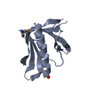


 PDBj
PDBj Assembly


 Mass: 92.094 Da / Num. of mol.: 1 / Source method: obtained synthetically / Formula: C3H8O3
 Mass: 18.015 Da / Num. of mol.: 37 / Source method: isolated from a natural source / Formula: H2O
 Sample preparation
 / Beamline: X29A / Wavelength: 0.9791 Å
 Processing