- PDB-3nqw: A metazoan ortholog of SpoT hydrolyzes ppGpp and plays a role in ... -
+
Open data
ID or keywords:
Loading...
-
Basic information
Entry
Database: PDB / ID: 3nqw
Title
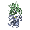
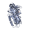
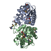
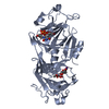
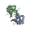
A metazoan ortholog of SpoT hydrolyzes ppGpp and plays a role in starvation responses
Components
CG11900
Keywords
HYDROLASE / stringent response / pyrophosphohydrolase / HD (Histidine and Aspartic acid) family / ppGpp hydrolase
Function / homology
Function and homology information
guanosine tetraphosphate catabolic process / guanosine-3',5'-bis(diphosphate) 3'-diphosphatase / guanosine-3',5'-bis(diphosphate) 3'-diphosphatase activity / response to starvation / metal ion binding Similarity search - Function
: / HD domain / Hypothetical protein af1432 / Hypothetical protein af1432 / HD domain profile. / HD domain / HD domain / Metal dependent phosphohydrolases with conserved 'HD' motif. / HD/PDEase domain / Orthogonal Bundle / Mainly Alpha Similarity search - Domain/homology
Protocol: SINGLE WAVELENGTH / Monochromatic (M) / Laue (L): M / Scattering type: x-ray
Radiation wavelength
Wavelength: 1.23985 Å / Relative weight: 1
Reflection
Redundancy: 3.5 % / Av σ(I) over netI: 20.02 / Number: 26676 / Rmerge(I) obs: 0.071 / Χ2: 1.31 / D res high: 2.9 Å / D res low: 50 Å / Num. obs: 7539 / % possible obs: 96.5
In the structure databanks used in Yorodumi, some data are registered as the other names, "COVID-19 virus" and "2019-nCoV". Here are the details of the virus and the list of structure data.
Jan 31, 2019. EMDB accession codes are about to change! (news from PDBe EMDB page)
EMDB accession codes are about to change! (news from PDBe EMDB page)
The allocation of 4 digits for EMDB accession codes will soon come to an end. Whilst these codes will remain in use, new EMDB accession codes will include an additional digit and will expand incrementally as the available range of codes is exhausted. The current 4-digit format prefixed with “EMD-” (i.e. EMD-XXXX) will advance to a 5-digit format (i.e. EMD-XXXXX), and so on. It is currently estimated that the 4-digit codes will be depleted around Spring 2019, at which point the 5-digit format will come into force.
The EM Navigator/Yorodumi systems omit the EMD- prefix.
Related info.:Q: What is EMD? / ID/Accession-code notation in Yorodumi/EM Navigator
Yorodumi is a browser for structure data from EMDB, PDB, SASBDB, etc.
This page is also the successor to EM Navigator detail page, and also detail information page/front-end page for Omokage search.
The word "yorodu" (or yorozu) is an old Japanese word meaning "ten thousand". "mi" (miru) is to see.
Related info.:EMDB / PDB / SASBDB / Comparison of 3 databanks / Yorodumi Search / Aug 31, 2016. New EM Navigator & Yorodumi / Yorodumi Papers / Jmol/JSmol / Function and homology information / Changes in new EM Navigator and Yorodumi
 Movie
Movie Controller

 Yorodumi
 Open data
 Basic information
 Components
 Keywords
 Function and homology information
 X-RAY DIFFRACTION /
  Authors
 Citation
 Structure visualization
 Downloads & links
 Other downloads
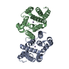


 PDBj
PDBj
 Assembly


 Mass: 54.938 Da / Num. of mol.: 2 / Source method: obtained synthetically / Formula: Mn
 Mass: 96.063 Da / Num. of mol.: 2 / Source method: obtained synthetically / Formula: SO4
 Mass: 18.015 Da / Num. of mol.: 36 / Source method: isolated from a natural source / Formula: H2O
 Sample preparation
 / Beamline: 6C1 / Wavelength: 1.23985 Å
 Processing