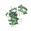
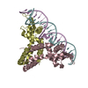
CRYSTAL PACKING AND SIZE EXCLUSION CHROMATOGRAPHY COUPLED WITH STATIC LIGHT SCATTERING ANALYSES SUPPORT THE ASSIGNMENT OF A DIMER AS A SIGNIFICANT OLIGOMERIZATION STATE IN SOLUTION.
-
Components
#1: Protein
MarR-familytranscriptionalregulator
Mass: 22458.258 Da / Num. of mol.: 2 Source method: isolated from a genetically manipulated source Source: (gene. exp.) Clostridium difficile (bacteria) / Strain: 630 / Gene: CD1569 / Plasmid: SpeedET / Production host: Escherichia coli (E. coli) / Strain (production host): HK100 / References: UniProt: Q186C2
Mass: 18.015 Da / Num. of mol.: 63 / Source method: isolated from a natural source / Formula: H2O
Has protein modification
Y
Sequence details
THIS CONSTRUCT (1-188) WAS EXPRESSED WITH THE PURIFICATION TAG MGSDKIHHHHHHENLYFQG. THE TAG WAS ...THIS CONSTRUCT (1-188) WAS EXPRESSED WITH THE PURIFICATION TAG MGSDKIHHHHHHENLYFQG. THE TAG WAS REMOVED WITH TEV PROTEASE LEAVING ONLY A GLYCINE (0) FOLLOWED BY THE TARGET SEQUENCE.
-
Experimental details
-
Experiment
Experiment
Method: X-RAY DIFFRACTION / Number of used crystals: 1
-
Sample preparation
Crystal
Density Matthews: 2.15 Å3/Da / Density % sol: 42.68 %
Crystal grow
Temperature: 277 K / Method: vapor diffusion, sitting drop / pH: 7.47 Details: 47.0000% polyethylene glycol 200, 0.1M HEPES pH 7.47, NANODROP, VAPOR DIFFUSION, SITTING DROP, temperature 277K
Monochromator: Double crystal monochromator / Protocol: SINGLE WAVELENGTH / Monochromatic (M) / Laue (L): M / Scattering type: x-ray
Radiation wavelength
Wavelength: 0.97911 Å / Relative weight: 1
Reflection
Resolution: 2.2→29.672 Å / Num. obs: 20101 / % possible obs: 98.8 % / Observed criterion σ(I): -3 / Biso Wilson estimate: 42.65 Å2 / Rmerge(I) obs: 0.059 / Net I/σ(I): 12.61
Reflection shell
Resolution (Å)
Rmerge(I) obs
Mean I/σ(I) obs
Num. measured obs
Num. unique obs
% possible all
2.2-2.28
0.681
1.8
11021
3579
93.1
2.28-2.37
0.505
2.5
11870
3705
99.8
2.37-2.48
0.381
3.3
12421
3868
99.6
2.48-2.61
0.27
4.6
12055
3748
99.6
2.61-2.77
0.194
6.1
11933
3712
99.8
2.77-2.98
0.135
8.6
11959
3712
99.9
2.98-3.28
0.081
13.2
12383
3834
99.7
3.28-3.76
0.046
21
12374
3822
99.5
3.76-4.72
0.029
30.6
12082
3743
99.5
4.72-29.672
0.03
33.7
12033
3780
97.8
-
Phasing
Phasing
Method: SAD
-
Processing
Software
Name
Version
Classification
NB
REFMAC
5.5.0110
refinement
PHENIX
refinement
SOLVE
phasing
MolProbity
3beta29
modelbuilding
XSCALE
datascaling
PDB_EXTRACT
3.006
dataextraction
XDS
datareduction
Refinement
Method to determine structure: SAD / Resolution: 2.2→29.672 Å / Cor.coef. Fo:Fc: 0.95 / Cor.coef. Fo:Fc free: 0.932 / Occupancy max: 1 / Occupancy min: 0.15 / SU B: 10.926 / SU ML: 0.134 / Cross valid method: THROUGHOUT / σ(F): 0 / ESU R: 0.287 / ESU R Free: 0.211 Stereochemistry target values: MAXIMUM LIKELIHOOD WITH PHASES Details: 1. HYDROGENS HAVE BEEN ADDED IN THE RIDING POSITIONS. 2. ATOM RECORDS CONTAIN SUM OF TLS AND RESIDUAL B FACTORS. ANISOU RECORDS CONTAIN SUM OF TLS AND RESIDUAL U FACTORS. 3. WATERS WERE ...Details: 1. HYDROGENS HAVE BEEN ADDED IN THE RIDING POSITIONS. 2. ATOM RECORDS CONTAIN SUM OF TLS AND RESIDUAL B FACTORS. ANISOU RECORDS CONTAIN SUM OF TLS AND RESIDUAL U FACTORS. 3. WATERS WERE EXCLUDED FROM AUTOMATIC TLS ASSIGNMENT. 4. A MET-INHIBITION PROTOCOL WAS USED FOR SELENOMETHIONINE INCORPORATION DURING PROTEIN EXPRESSION. THE OCCUPANCY OF THE SE ATOMS IN THE MSE RESIDUES WAS REDUCED TO 0.75 TO ACCOUNT FOR THE REDUCED SCATTERING POWER DUE TO PARTIAL S-MET INCORPORATION. 5. RESIDUES 160-164 OF CHAIN A AND 155-163 OF CHAIN B WERE NOT MODELED DUE TO POOR ELECTRON DENSITY IN THOSE REGIONS. 6. SODIUM (NA) FROM THE PROTEIN BUFFER, AND GLYCEROL (GOL) AND POLYETHYLENE GLYCOL (PG4) FROM THE CRYSTALLIZATION/ CRYOPROTECTANT SOLUTIONS HAVE BEEN MODELED INTO THE SOLVENT STRUCTURE. 7. TLS GROUPS WERE ASSIGNED WITH THE AID OF THE TLSMD SERVER.
Rfactor
Num. reflection
% reflection
Selection details
Rfree
0.237
1021
5.1 %
RANDOM
Rwork
0.197
-
-
-
obs
0.199
20052
99.51 %
-
Solvent computation
Ion probe radii: 0.8 Å / Shrinkage radii: 0.8 Å / VDW probe radii: 1.4 Å / Solvent model: MASK
In the structure databanks used in Yorodumi, some data are registered as the other names, "COVID-19 virus" and "2019-nCoV". Here are the details of the virus and the list of structure data.
Jan 31, 2019. EMDB accession codes are about to change! (news from PDBe EMDB page)
EMDB accession codes are about to change! (news from PDBe EMDB page)
The allocation of 4 digits for EMDB accession codes will soon come to an end. Whilst these codes will remain in use, new EMDB accession codes will include an additional digit and will expand incrementally as the available range of codes is exhausted. The current 4-digit format prefixed with “EMD-” (i.e. EMD-XXXX) will advance to a 5-digit format (i.e. EMD-XXXXX), and so on. It is currently estimated that the 4-digit codes will be depleted around Spring 2019, at which point the 5-digit format will come into force.
The EM Navigator/Yorodumi systems omit the EMD- prefix.
Related info.:Q: What is EMD? / ID/Accession-code notation in Yorodumi/EM Navigator
Yorodumi is a browser for structure data from EMDB, PDB, SASBDB, etc.
This page is also the successor to EM Navigator detail page, and also detail information page/front-end page for Omokage search.
The word "yorodu" (or yorozu) is an old Japanese word meaning "ten thousand". "mi" (miru) is to see.
Related info.:EMDB / PDB / SASBDB / Comparison of 3 databanks / Yorodumi Search / Aug 31, 2016. New EM Navigator & Yorodumi / Yorodumi Papers / Jmol/JSmol / Function and homology information / Changes in new EM Navigator and Yorodumi
 Movie
Movie Controller

 Yorodumi
 Open data
 Basic information
 Components
 Keywords
 Function and homology information
 Clostridium difficile (bacteria)
 X-RAY DIFFRACTION /
  Authors
 Citation
 Structure visualization
 Downloads & links
 Other downloads


 PDBj
PDBj Assembly
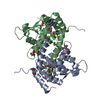

 Mass: 92.094 Da / Num. of mol.: 2 / Source method: obtained synthetically / Formula: C3H8O3
 Mass: 194.226 Da / Num. of mol.: 5 / Source method: obtained synthetically / Formula: C8H18O5 / Comment: precipitant*YM
 Mass: 22.990 Da / Num. of mol.: 1 / Source method: obtained synthetically / Formula: Na
 Mass: 18.015 Da / Num. of mol.: 63 / Source method: isolated from a natural source / Formula: H2O
 Sample preparation
 / Beamline: BL9-2 / Wavelength: 0.97911
  Processing