 Movie
Movie Controller

[English] 日本語
 Yorodumi
- PDB-3nou: Light-induced intermediate structure L3 of P. aeruginosa bacterio... -
+ Open data

- Basic information
| Entry | Database: PDB / ID: 3nou | ||||||
|---|---|---|---|---|---|---|---|
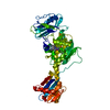
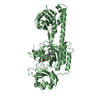
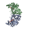
| Title | Light-induced intermediate structure L3 of P. aeruginosa bacteriophytochrome | ||||||
 Components Components | Bacteriophytochrome | ||||||
 Keywords Keywords | SIGNALING PROTEIN / Intermediate structure | ||||||
| Function / homology |  Function and homology information Function and homology informationosmosensory signaling via phosphorelay pathway / detection of visible light / phosphorelay response regulator activity / phosphorelay sensor kinase activity / histidine kinase / photoreceptor activity / protein kinase activator activity / regulation of DNA-templated transcription / ATP binding / identical protein binding Similarity search - Function | ||||||
| Biological species |   Pseudomonas aeruginosa (bacteria) Pseudomonas aeruginosa (bacteria) | ||||||
| Method |  X-RAY DIFFRACTION / SYNCHROTRON / difference Fourier Method / Resolution: 3 Å X-RAY DIFFRACTION / SYNCHROTRON / difference Fourier Method / Resolution: 3 Å | ||||||
 Authors Authors | Yang, X. / Ren, Z. / Moffat, K. | ||||||
 Citation Citation | Journal: Nature / Year: 2011 Title: Temperature-scan cryocrystallography reveals reaction intermediates in bacteriophytochrome. Authors: Yang, X. / Ren, Z. / Kuk, J. / Moffat, K. | ||||||
| History |
|
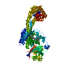
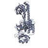
- Structure visualization
| Structure viewer | Molecule: MolmilJmol/JSmol |
|---|
- Downloads & links
-Download
| PDBx/mmCIF format | 3nou.cif.gz | 108.5 KB | Display | PDBx/mmCIF format |
|---|---|---|---|---|
| PDB format | pdb3nou.ent.gz | 83.8 KB | Display | PDB format |
| PDBx/mmJSON format | 3nou.json.gz | Tree view | PDBx/mmJSON format | |
| Others |  Other downloads Other downloads |
-Validation report
| Arichive directory | https://data.pdbj.org/pub/pdb/validation_reports/no/3nouftp://data.pdbj.org/pub/pdb/validation_reports/no/3nou | HTTPS FTP |
|---|
-Related structure data
| Related structure data |  3nhqSC  3nopC  3notC S: Starting model for refinement C: citing same article ( |
|---|---|
| Similar structure data |


-Links
 PDBj
PDBj


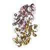
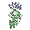
- Assembly
| Deposited unit | 
| ||||||||||||||||||||||||||||||||||||
|---|---|---|---|---|---|---|---|---|---|---|---|---|---|---|---|---|---|---|---|---|---|---|---|---|---|---|---|---|---|---|---|---|---|---|---|---|---|
| 1 |
| ||||||||||||||||||||||||||||||||||||
| Unit cell |
| ||||||||||||||||||||||||||||||||||||
| Noncrystallographic symmetry (NCS) | NCS oper:
|
-Components
| #1: Protein | Mass: 56823.230 Da / Num. of mol.: 1 / Fragment: Photosensory Core Module Source method: isolated from a genetically manipulated source Source: (gene. exp.) Pseudomonas aeruginosa (bacteria) / Strain: PA01 / Gene: bphP, PA4117 / Plasmid: pET24 / Production host: |
|---|---|
| #2: Chemical | ChemComp-BLA /   Mass: 582.646 Da / Num. of mol.: 1 / Source method: obtained synthetically / Formula: C33H34N4O6 Mass: 582.646 Da / Num. of mol.: 1 / Source method: obtained synthetically / Formula: C33H34N4O6 |
| Has protein modification | Y |
-Experimental details
-Experiment
| Experiment | Method: X-RAY DIFFRACTION / Number of used crystals: 6 |
|---|
- Sample preparation
| Crystal | Density Matthews: 3.03 Å3/Da / Density % sol: 59.4 % |
|---|---|
| Crystal grow | Temperature: 293 K / Method: vapor diffusion / pH: 7.7 Details: 10mg/ml protein, 0.45M ammonium phosphate, 0.1M Tris buffer, pH 7.7, VAPOR DIFFUSION, temperature 293K |
-Data collection
| Diffraction |
| |||||||||
|---|---|---|---|---|---|---|---|---|---|---|
| Diffraction source | Source: SYNCHROTRON / Site: APS  / Beamline: 21-ID-G / Wavelength: 0.97857 Å / Beamline: 21-ID-G / Wavelength: 0.97857 Å | |||||||||
| Detector | Type: RAYONIX MX-300 / Detector: CCD / Date: Nov 12, 2008 | |||||||||
| Radiation | Monochromator: C(111) / Protocol: SINGLE WAVELENGTH / Monochromatic (M) / Laue (L): M / Scattering type: x-ray | |||||||||
| Radiation wavelength | Wavelength: 0.97857 Å / Relative weight: 1 | |||||||||
| Reflection | Resolution: 2.9→50 Å / Num. all: 134289 / Num. obs: 132947 / % possible obs: 99 % / Redundancy: 5.6 % / Rmerge(I) obs: 0.106 |
- Processing
| Software |
| ||||||||||||
|---|---|---|---|---|---|---|---|---|---|---|---|---|---|
| Refinement | Method to determine structure: difference Fourier Method Starting model: PDB ENTRY 3NHQ Resolution: 3→50 Å / σ(F): 0 / σ(I): 0 Details: This cryo-trapped structure was determined based on difference Fourier method. The L1 (3NOP), L2(3NOT) and L3(3NOU) structures were refined jointly in real space against a set of (Flight- ...Details: This cryo-trapped structure was determined based on difference Fourier method. The L1 (3NOP), L2(3NOT) and L3(3NOU) structures were refined jointly in real space against a set of (Flight-Fdark) difference maps representing mixtures of the L1, L2 and L3 structures in variable relative concentrations using software DynamiX. DynamiX is a collection of software tools for analyzing dynamic crystallographic data developed by Zhong Ren. Algorithms and methods are described in Ren, Z et al. Resolution of structural heterogeneity in dynamic and static crystallography. Manuscript in preparation.
| ||||||||||||
| Refinement step | Cycle: LAST / Resolution: 3→50 Å
|
