 Movie
Movie Controller

[English] 日本語
 Yorodumi
- PDB-3nkg: Crystal Structure of GEBA250068378 from Sulfurospirillum deleyianum -
+ Open data

- Basic information
| Entry | Database: PDB / ID: 3nkg | ||||||
|---|---|---|---|---|---|---|---|
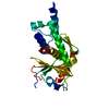
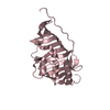
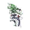
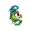
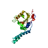
| Title | Crystal Structure of GEBA250068378 from Sulfurospirillum deleyianum | ||||||
 Components Components | uncharacterized protein GEBA250068378 | ||||||
 Keywords Keywords | structural genomics / unknown function / GEBA target / bata-fold / PSI-2 / Protein Structure Initiative / Midwest Center for Structural Genomics / MCSG | ||||||
| Function / homology | Jelly Rolls - #790 / Domain of unknown function DUF5625 / Domain of unknown function (DUF5625) / Jelly Rolls / Sandwich / Mainly Beta / ACETIC ACID / DUF5625 domain-containing protein Function and homology information Function and homology information | ||||||
| Biological species |  Sulfurospirillum deleyianum (bacteria) Sulfurospirillum deleyianum (bacteria) | ||||||
| Method |  X-RAY DIFFRACTION / SYNCHROTRON / SAD / Resolution: 2 Å X-RAY DIFFRACTION / SYNCHROTRON / SAD / Resolution: 2 Å | ||||||
 Authors Authors | Kim, Y. / Marshall, N. / Cobb, G. / Eisen, J. / Kerfeld, C. / Joachimiak, A. / Midwest Center for Structural Genomics (MCSG) | ||||||
 Citation Citation | Journal: To be Published / Year: 2010 Title: Crystal Structure of GEBA250068378 from Sulfurospirillum deleyianum Authors: Kim, Y. / Marshall, N. / Cobb, G. / Eisen, J. / Kerfeld, C. / Joachimiak, A. | ||||||
| History |
|
- Structure visualization
| Structure viewer | Molecule: MolmilJmol/JSmol |
|---|
- Downloads & links
-Download
| PDBx/mmCIF format | 3nkg.cif.gz | 159 KB | Display | PDBx/mmCIF format |
|---|---|---|---|---|
| PDB format | pdb3nkg.ent.gz | 125.8 KB | Display | PDB format |
| PDBx/mmJSON format | 3nkg.json.gz | Tree view | PDBx/mmJSON format | |
| Others |  Other downloads Other downloads |
-Validation report
| Arichive directory | https://data.pdbj.org/pub/pdb/validation_reports/nk/3nkgftp://data.pdbj.org/pub/pdb/validation_reports/nk/3nkg | HTTPS FTP |
|---|
-Related structure data
| Similar structure data | |
|---|---|
| Other databases |


-Links
 PDBj
PDBj

- Assembly
| Deposited unit | 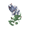
| ||||||||
|---|---|---|---|---|---|---|---|---|---|
| 1 | 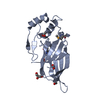
| ||||||||
| 2 | 
| ||||||||
| Unit cell |
|
-Components
| #1: Protein | Mass: 19891.121 Da / Num. of mol.: 2 Source method: isolated from a genetically manipulated source Source: (gene. exp.) Sulfurospirillum deleyianum (bacteria) / Strain: DSM 6946 / Gene: Sdel_1248 / Plasmid: pMCSG7 / Production host: #2: Chemical | ChemComp-ACY /   Mass: 60.052 Da / Num. of mol.: 5 / Source method: obtained synthetically / Formula: C2H4O2 Mass: 60.052 Da / Num. of mol.: 5 / Source method: obtained synthetically / Formula: C2H4O2#3: Chemical |   Mass: 92.094 Da / Num. of mol.: 3 / Source method: obtained synthetically / Formula: C3H8O3 Mass: 92.094 Da / Num. of mol.: 3 / Source method: obtained synthetically / Formula: C3H8O3#4: Water | ChemComp-HOH / |  Mass: 18.015 Da / Num. of mol.: 294 / Source method: isolated from a natural source / Formula: H2O Mass: 18.015 Da / Num. of mol.: 294 / Source method: isolated from a natural source / Formula: H2OHas protein modification | Y | |
|---|
-Experimental details
-Experiment
| Experiment | Method: X-RAY DIFFRACTION / Number of used crystals: 1 |
|---|
- Sample preparation
| Crystal | Density Matthews: 2.33 Å3/Da / Density % sol: 47.24 % |
|---|---|
| Crystal grow | Temperature: 289 K / Method: vapor diffusion, sitting drop / pH: 6.5 Details: 0.16 M Magnesium Acetate 0.08 M Sodium Cacodylate pH 6.5, 16% PEG 8000, 20% Glycerol, VAPOR DIFFUSION, SITTING DROP, temperature 289K |
-Data collection
| Diffraction | Mean temperature: 100 K |
|---|---|
| Diffraction source | Source: SYNCHROTRON / Site: APS  / Beamline: 19-ID / Wavelength: 0.97935 Å / Beamline: 19-ID / Wavelength: 0.97935 Å |
| Detector | Type: ADSC QUANTUM 315r / Detector: CCD / Date: Jun 21, 2009 / Details: mirrors |
| Radiation | Monochromator: double crystal monochromator / Protocol: SINGLE WAVELENGTH / Monochromatic (M) / Laue (L): M / Scattering type: x-ray |
| Radiation wavelength | Wavelength: 0.97935 Å / Relative weight: 1 |
| Reflection | Resolution: 2→50 Å / Num. all: 25082 / Num. obs: 25082 / % possible obs: 99.7 % / Observed criterion σ(F): 0 / Observed criterion σ(I): 0 / Redundancy: 4.4 % / Biso Wilson estimate: 24.61 Å2 / Rsym value: 0.086 / Net I/σ(I): 9.9 |
- Processing
| Software |
| |||||||||||||||||||||||||||||||||||||||||||||||||||||||||||||||||||||||||||||
|---|---|---|---|---|---|---|---|---|---|---|---|---|---|---|---|---|---|---|---|---|---|---|---|---|---|---|---|---|---|---|---|---|---|---|---|---|---|---|---|---|---|---|---|---|---|---|---|---|---|---|---|---|---|---|---|---|---|---|---|---|---|---|---|---|---|---|---|---|---|---|---|---|---|---|---|---|---|---|
| Refinement | Method to determine structure: SAD / Resolution: 2→44.087 Å / SU ML: 0.2 / Isotropic thermal model: mixed / Cross valid method: THROUGHOUT / σ(F): 0 / Stereochemistry target values: MLHL
| |||||||||||||||||||||||||||||||||||||||||||||||||||||||||||||||||||||||||||||
| Solvent computation | Shrinkage radii: 0.9 Å / VDW probe radii: 1.11 Å / Solvent model: FLAT BULK SOLVENT MODEL / Bsol: 40.507 Å2 / ksol: 0.351 e/Å3 | |||||||||||||||||||||||||||||||||||||||||||||||||||||||||||||||||||||||||||||
| Displacement parameters | Biso mean: 35.1 Å2
| |||||||||||||||||||||||||||||||||||||||||||||||||||||||||||||||||||||||||||||
| Refinement step | Cycle: LAST / Resolution: 2→44.087 Å
| |||||||||||||||||||||||||||||||||||||||||||||||||||||||||||||||||||||||||||||
| Refine LS restraints |
| |||||||||||||||||||||||||||||||||||||||||||||||||||||||||||||||||||||||||||||
| LS refinement shell | Refine-ID: X-RAY DIFFRACTION
| |||||||||||||||||||||||||||||||||||||||||||||||||||||||||||||||||||||||||||||
| Refinement TLS params. | Method: refined / Refine-ID: X-RAY DIFFRACTION
| |||||||||||||||||||||||||||||||||||||||||||||||||||||||||||||||||||||||||||||
| Refinement TLS group |
|
