 Movie
Movie Controller

[English] 日本語
 Yorodumi
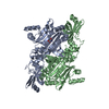
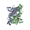
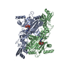
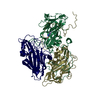
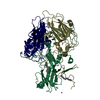
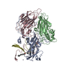
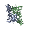
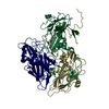
- PDB-3nen: Unliganded aspartyl-tRNA synthetase from thermococcus kodakarensis -
+ Open data

- Basic information
| Entry | Database: PDB / ID: 3nen | ||||||
|---|---|---|---|---|---|---|---|
| Title | Unliganded aspartyl-tRNA synthetase from thermococcus kodakarensis | ||||||
 Components Components | Aspartyl-tRNA synthetase | ||||||
 Keywords Keywords | LIGASE / aminoacyl-tRNA synthetase / Rossmann fold Ob fold / Aspartic acid / ATP-Mg / tRNA | ||||||
| Function / homology |  Function and homology information Function and homology informationaspartate-tRNA ligase / aspartate-tRNA ligase activity / aspartyl-tRNA aminoacylation / aminoacyl-tRNA synthetase multienzyme complex / magnesium ion binding / RNA binding / ATP binding / cytosol Similarity search - Function | ||||||
| Biological species |   Thermococcus kodakarensis (archaea) Thermococcus kodakarensis (archaea) | ||||||
| Method |  X-RAY DIFFRACTION / SYNCHROTRON / MOLECULAR REPLACEMENT / Resolution: 2.4 Å X-RAY DIFFRACTION / SYNCHROTRON / MOLECULAR REPLACEMENT / Resolution: 2.4 Å | ||||||
 Authors Authors | Schmitt, E. / Moras, D. / Moulinier, L. | ||||||
 Citation Citation | Journal: Embo J. / Year: 1998 Title: Crystal structure of aspartyl-tRNA synthetase from Pyrococcus kodakaraensis KOD: archaeon specificity and catalytic mechanism of adenylate formation Authors: Schmitt, E. / Moulinier, L. / Fujiwara, S. / Imanaka, T. / Thierry, J.C. / Moras, D. | ||||||
| History |
|
- Structure visualization
| Structure viewer | Molecule: MolmilJmol/JSmol |
|---|
- Downloads & links
-Download
| PDBx/mmCIF format | 3nen.cif.gz | 183.1 KB | Display | PDBx/mmCIF format |
|---|---|---|---|---|
| PDB format | pdb3nen.ent.gz | 147.5 KB | Display | PDB format |
| PDBx/mmJSON format | 3nen.json.gz | Tree view | PDBx/mmJSON format | |
| Others |  Other downloads Other downloads |
-Validation report
| Summary document | 3nen_validation.pdf.gz | 441.8 KB | Display | wwPDB validaton report |
|---|---|---|---|---|
| Full document | 3nen_full_validation.pdf.gz | 462.8 KB | Display | |
| Data in XML | 3nen_validation.xml.gz | 33.1 KB | Display | |
| Data in CIF | 3nen_validation.cif.gz | 44.9 KB | Display | |
| Arichive directory | https://data.pdbj.org/pub/pdb/validation_reports/ne/3nenftp://data.pdbj.org/pub/pdb/validation_reports/ne/3nen | HTTPS FTP |
-Related structure data
| Related structure data |  1b8aC  3nelC  3nemC 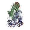 1aszS C: citing same article ( S: Starting model for refinement |
|---|---|
| Similar structure data |

-Links
 PDBj
PDBj

- Assembly
| Deposited unit | 
| ||||||||
|---|---|---|---|---|---|---|---|---|---|
| 1 |
| ||||||||
| Unit cell |
|
-Components
| #1: Protein | Mass: 50979.414 Da / Num. of mol.: 2 Source method: isolated from a genetically manipulated source Source: (gene. exp.) Thermococcus kodakarensis (archaea) / Gene: aspS, TK0492 / Production host:  #2: Water | ChemComp-HOH / |  Mass: 18.015 Da / Num. of mol.: 77 / Source method: isolated from a natural source / Formula: H2O Mass: 18.015 Da / Num. of mol.: 77 / Source method: isolated from a natural source / Formula: H2O |
|---|
-Experimental details
-Experiment
| Experiment | Method: X-RAY DIFFRACTION / Number of used crystals: 1 |
|---|
- Sample preparation
| Crystal | Density Matthews: 3.32 Å3/Da / Density % sol: 62.99 % |
|---|---|
| Crystal grow | Temperature: 297 K Details: peg and ethylen glycol, VAPOR DIFFUSION, HANGING DROP, temperature 297K |
-Data collection
| Diffraction | Mean temperature: 120 K |
|---|---|
| Diffraction source | Source: SYNCHROTRON / Site: ESRF  / Beamline: BM30A / Wavelength: 0.98 / Beamline: BM30A / Wavelength: 0.98 |
| Detector | Type: MAR CCD 165 mm / Detector: CCD / Date: Dec 10, 1996 |
| Radiation | Monochromator: DOUBLE CRYSTAL MONOCHROMATOR / Protocol: SINGLE WAVELENGTH / Monochromatic (M) / Laue (L): M / Scattering type: x-ray |
| Radiation wavelength | Wavelength: 0.98 Å / Relative weight: 1 |
| Reflection | Resolution: 2.4→37.268 Å / Num. obs: 48410 / % possible obs: 89.9 % / Observed criterion σ(I): 0 / Redundancy: 5.7 % / Biso Wilson estimate: 48.8 Å2 / Rsym value: 0.099 |
- Processing
| Software |
| ||||||||||||||||||||||||||||||||||||||||||||||||||||||||||||
|---|---|---|---|---|---|---|---|---|---|---|---|---|---|---|---|---|---|---|---|---|---|---|---|---|---|---|---|---|---|---|---|---|---|---|---|---|---|---|---|---|---|---|---|---|---|---|---|---|---|---|---|---|---|---|---|---|---|---|---|---|---|
| Refinement | Method to determine structure: MOLECULAR REPLACEMENT Starting model: PDB ENTRY 1ASZ Resolution: 2.4→37.27 Å / Cross valid method: THROUGHOUT / σ(F): 2 / Stereochemistry target values: Engh & Huber
| ||||||||||||||||||||||||||||||||||||||||||||||||||||||||||||
| Displacement parameters | Biso mean: 38.1 Å2 | ||||||||||||||||||||||||||||||||||||||||||||||||||||||||||||
| Refinement step | Cycle: LAST / Resolution: 2.4→37.27 Å
| ||||||||||||||||||||||||||||||||||||||||||||||||||||||||||||
| Refine LS restraints |
|
