 Movie
Movie Controller

[English] 日本語
 Yorodumi
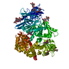
- PDB-3ndv: Crystal structure of the N-terminal beta-aminopeptidase BapA in c... -
+ Open data

- Basic information
| Entry | Database: PDB / ID: 3ndv | ||||||
|---|---|---|---|---|---|---|---|
| Title | Crystal structure of the N-terminal beta-aminopeptidase BapA in complex with ampicillin | ||||||
 Components Components | Beta-peptidyl aminopeptidase | ||||||
 Keywords Keywords | HYDROLASE/ANTIBIOTIC / Ntn-hydrolase / beta-aminopeptidase / beta-peptide / alpha-beta-beta-alpha sandwich / N-terminal beta-aminopeptidase / HYDROLASE-ANTIBIOTIC complex | ||||||
| Function / homology |  Function and homology information Function and homology informationbeta-peptidyl aminopeptidase / aminopeptidase activity / periplasmic space / proteolysis / identical protein binding Similarity search - Function | ||||||
| Biological species |  Sphingosinicella xenopeptidilytica (bacteria) Sphingosinicella xenopeptidilytica (bacteria) | ||||||
| Method |  X-RAY DIFFRACTION / SYNCHROTRON / MOLECULAR REPLACEMENT / Resolution: 1.7 Å X-RAY DIFFRACTION / SYNCHROTRON / MOLECULAR REPLACEMENT / Resolution: 1.7 Å | ||||||
 Authors Authors | Merz, T. / Heck, T. / Geueke, B. / Kohler, H.-P.E. / Gruetter, M.G. | ||||||
 Citation Citation | Journal: To be Published Title: Crystal structures and inhibition of the beta-aminopeptidase BapA, a new ampicillin-recognizing member of the N-terminal nucleophile hydrolase family Authors: Heck, T. / Merz, T. / Geueke, B. / Gruetter, M.G. / Kohler, H.-P.E. | ||||||
| History |
|
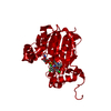
- Structure visualization
| Structure viewer | Molecule: MolmilJmol/JSmol |
|---|
- Downloads & links
-Download
| PDBx/mmCIF format | 3ndv.cif.gz | 299.8 KB | Display | PDBx/mmCIF format |
|---|---|---|---|---|
| PDB format | pdb3ndv.ent.gz | 241.9 KB | Display | PDB format |
| PDBx/mmJSON format | 3ndv.json.gz | Tree view | PDBx/mmJSON format | |
| Others |  Other downloads Other downloads |
-Validation report
| Arichive directory | https://data.pdbj.org/pub/pdb/validation_reports/nd/3ndvftp://data.pdbj.org/pub/pdb/validation_reports/nd/3ndv | HTTPS FTP |
|---|
-Related structure data
| Related structure data | 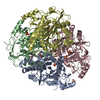 3n2wS S: Starting model for refinement |
|---|---|
| Similar structure data |


-Links
 PDBj
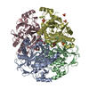
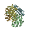
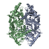
PDBj- Assembly
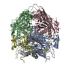
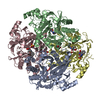
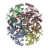
| Deposited unit | 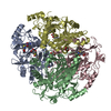
| ||||||||
|---|---|---|---|---|---|---|---|---|---|
| 1 |
| ||||||||
| Unit cell |
|
-Components
| #1: Protein | Mass: 38634.836 Da / Num. of mol.: 4 Source method: isolated from a genetically manipulated source Source: (gene. exp.) Sphingosinicella xenopeptidilytica (bacteria)Strain: 3-2W4 / Gene: bapA / Plasmid: pYBapA / Production host: #2: Chemical | ChemComp-GOL /   Mass: 92.094 Da / Num. of mol.: 4 / Source method: obtained synthetically / Formula: C3H8O3 Mass: 92.094 Da / Num. of mol.: 4 / Source method: obtained synthetically / Formula: C3H8O3#3: Chemical | ChemComp-AIC / (   Mass: 349.405 Da / Num. of mol.: 4 / Source method: obtained synthetically / Formula: C16H19N3O4S / Comment: antibiotic*YM Mass: 349.405 Da / Num. of mol.: 4 / Source method: obtained synthetically / Formula: C16H19N3O4S / Comment: antibiotic*YM#4: Chemical | 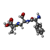  Mass: 351.421 Da / Num. of mol.: 2 / Source method: obtained synthetically / Formula: C16H21N3O4S Mass: 351.421 Da / Num. of mol.: 2 / Source method: obtained synthetically / Formula: C16H21N3O4S#5: Water | ChemComp-HOH / |  Mass: 18.015 Da / Num. of mol.: 1138 / Source method: isolated from a natural source / Formula: H2O Mass: 18.015 Da / Num. of mol.: 1138 / Source method: isolated from a natural source / Formula: H2O |
|---|
-Experimental details
-Experiment
| Experiment | Method: X-RAY DIFFRACTION / Number of used crystals: 1 |
|---|
- Sample preparation
| Crystal | Density Matthews: 2.61 Å3/Da / Density % sol: 52.88 % |
|---|---|
| Crystal grow | Temperature: 295 K / Method: vapor diffusion, sitting drop / pH: 7.5 Details: 15mg/ml protein, 1.5M ammonium sulfate, 0.1M HEPES pH 7.5, VAPOR DIFFUSION, SITTING DROP, temperature 295K |
-Data collection
| Diffraction | Mean temperature: 100 K |
|---|---|
| Diffraction source | Source: SYNCHROTRON / Site: SLS  / Beamline: X06SA / Wavelength: 1 Å / Beamline: X06SA / Wavelength: 1 Å |
| Detector | Type: PSI PILATUS 6M / Detector: PIXEL / Date: Feb 12, 2009 / Details: dynamically bendable mirror |
| Radiation | Monochromator: LN2 cooled fixed exit Si(111) monochromator / Protocol: SINGLE WAVELENGTH / Monochromatic (M) / Laue (L): M / Scattering type: x-ray |
| Radiation wavelength | Wavelength: 1 Å / Relative weight: 1 |
| Reflection | Resolution: 1.7→50 Å / Num. obs: 172088 / % possible obs: 98.7 % / Observed criterion σ(I): 3 / Redundancy: 3.31 % / Rmerge(I) obs: 0.16 / Rsym value: 0.116 / Net I/σ(I): 9.23 |
| Reflection shell | Resolution: 1.7→1.8 Å / Redundancy: 2.72 % / Rmerge(I) obs: 0.67 / Mean I/σ(I) obs: 2.2 / Num. unique all: 70374 / Rsym value: 0.581 / % possible all: 94.4 |
- Processing
| Software |
| |||||||||||||||||||||||||||||||||||||||||||||||||||||||||||||||||||||||||||||||||||||||||||||||||||||||||||||||||||||||||||||||||||||||||||||||||||||||||||||||||||||||||||||||||||||||||||||||||||||||||||||||||||||||||
|---|---|---|---|---|---|---|---|---|---|---|---|---|---|---|---|---|---|---|---|---|---|---|---|---|---|---|---|---|---|---|---|---|---|---|---|---|---|---|---|---|---|---|---|---|---|---|---|---|---|---|---|---|---|---|---|---|---|---|---|---|---|---|---|---|---|---|---|---|---|---|---|---|---|---|---|---|---|---|---|---|---|---|---|---|---|---|---|---|---|---|---|---|---|---|---|---|---|---|---|---|---|---|---|---|---|---|---|---|---|---|---|---|---|---|---|---|---|---|---|---|---|---|---|---|---|---|---|---|---|---|---|---|---|---|---|---|---|---|---|---|---|---|---|---|---|---|---|---|---|---|---|---|---|---|---|---|---|---|---|---|---|---|---|---|---|---|---|---|---|---|---|---|---|---|---|---|---|---|---|---|---|---|---|---|---|---|---|---|---|---|---|---|---|---|---|---|---|---|---|---|---|---|---|---|---|---|---|---|---|---|---|---|---|---|---|---|---|---|
| Refinement | Method to determine structure: MOLECULAR REPLACEMENT Starting model: PDB ENTRY 3N2W Resolution: 1.7→48.835 Å / SU ML: 0.18 / Isotropic thermal model: ISOTROPIC / Cross valid method: THROUGHOUT / σ(F): 1.99 / Stereochemistry target values: ML
| |||||||||||||||||||||||||||||||||||||||||||||||||||||||||||||||||||||||||||||||||||||||||||||||||||||||||||||||||||||||||||||||||||||||||||||||||||||||||||||||||||||||||||||||||||||||||||||||||||||||||||||||||||||||||
| Solvent computation | Shrinkage radii: 0.9 Å / VDW probe radii: 1.11 Å / Solvent model: FLAT BULK SOLVENT MODEL / Bsol: 60.428 Å2 / ksol: 0.4 e/Å3 | |||||||||||||||||||||||||||||||||||||||||||||||||||||||||||||||||||||||||||||||||||||||||||||||||||||||||||||||||||||||||||||||||||||||||||||||||||||||||||||||||||||||||||||||||||||||||||||||||||||||||||||||||||||||||
| Displacement parameters |
| |||||||||||||||||||||||||||||||||||||||||||||||||||||||||||||||||||||||||||||||||||||||||||||||||||||||||||||||||||||||||||||||||||||||||||||||||||||||||||||||||||||||||||||||||||||||||||||||||||||||||||||||||||||||||
| Refinement step | Cycle: LAST / Resolution: 1.7→48.835 Å
| |||||||||||||||||||||||||||||||||||||||||||||||||||||||||||||||||||||||||||||||||||||||||||||||||||||||||||||||||||||||||||||||||||||||||||||||||||||||||||||||||||||||||||||||||||||||||||||||||||||||||||||||||||||||||
| Refine LS restraints |
| |||||||||||||||||||||||||||||||||||||||||||||||||||||||||||||||||||||||||||||||||||||||||||||||||||||||||||||||||||||||||||||||||||||||||||||||||||||||||||||||||||||||||||||||||||||||||||||||||||||||||||||||||||||||||
| LS refinement shell |
|
