| 登録情報 | データベース: PDB / ID: 3ndp
|
|---|
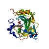
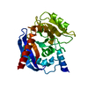
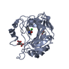
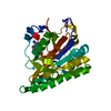
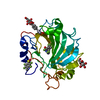
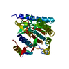
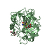
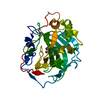
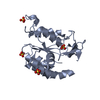
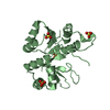
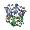
| タイトル | Crystal structure of human AK4(L171P) |
|---|
 要素 要素 | Adenylate kinase isoenzyme 4 |
|---|
 キーワード キーワード | TRANSFERASE / PARALLEL BETA-SHEET / ALPHA-HELICES |
|---|
| 機能・相同性 |  機能・相同性情報 機能・相同性情報
ribonucleoside diphosphate biosynthetic process / nucleoside triphosphate adenylate kinase activity / nucleoside-phosphate kinase / nucleoside monophosphate kinase activity / dAMP kinase activity / CMP kinase activity / dCMP kinase activity / AMP metabolic process / ADP biosynthetic process / nucleoside triphosphate biosynthetic process ...ribonucleoside diphosphate biosynthetic process / nucleoside triphosphate adenylate kinase activity / nucleoside-phosphate kinase / nucleoside monophosphate kinase activity / dAMP kinase activity / CMP kinase activity / dCMP kinase activity / AMP metabolic process / ADP biosynthetic process / nucleoside triphosphate biosynthetic process / regulation of oxidative phosphorylation / AMP kinase activity / nucleoside-diphosphate kinase / nucleobase-containing small molecule interconversion / Interconversion of nucleotide di- and triphosphates / GTP metabolic process / nucleoside diphosphate kinase activity / ATP metabolic process / cellular response to hypoxia / mitochondrial matrix / GTP binding / mitochondrion / ATP binding / cytoplasm類似検索 - 分子機能 Adenylate kinase 4, mitochondrial / Adenylate kinase 3/4, mitochondrial / Adenylate kinase, active site lid domain superfamily / Adenylate kinase, active site lid domain / Adenylate kinase, active site lid / Adenylate kinase subfamily / Adenylate kinase, conserved site / Adenylate kinase signature. / Adenylate kinase/UMP-CMP kinase / Adenylate kinase ...Adenylate kinase 4, mitochondrial / Adenylate kinase 3/4, mitochondrial / Adenylate kinase, active site lid domain superfamily / Adenylate kinase, active site lid domain / Adenylate kinase, active site lid / Adenylate kinase subfamily / Adenylate kinase, conserved site / Adenylate kinase signature. / Adenylate kinase/UMP-CMP kinase / Adenylate kinase / P-loop containing nucleotide triphosphate hydrolases / Rossmann fold / P-loop containing nucleoside triphosphate hydrolase / 3-Layer(aba) Sandwich / Alpha Beta類似検索 - ドメイン・相同性 |
|---|
| 生物種 |  Homo sapiens (ヒト) Homo sapiens (ヒト) |
|---|
| 手法 |  X線回折 / シンクロトロン / 分子置換 / 解像度: 2.3 Å X線回折 / シンクロトロン / 分子置換 / 解像度: 2.3 Å |
|---|
 データ登録者 データ登録者 | Liu, R. / Wang, Y. / Wei, Z. / Gong, W. |
|---|
 引用 引用 | ジャーナル: Biochem.Biophys.Res.Commun. / 年: 2009
タイトル: Crystal structure of human adenylate kinase 4 (L171P) suggests the role of hinge region in protein domain motion
著者: Liu, R. / Xu, H. / Wei, Z. / Wang, Y. / Lin, Y. / Gong, W. |
|---|
| 履歴 | | 登録 | 2010年6月7日 | 登録サイト: RCSB / 処理サイト: PDBJ |
|---|
| 改定 1.0 | 2010年6月23日 | Provider: repository / タイプ: Initial release |
|---|
| 改定 1.1 | 2011年7月13日 | Group: Version format compliance |
|---|
| 改定 1.2 | 2017年11月8日 | Group: Refinement description / カテゴリ: software |
|---|
| 改定 1.3 | 2024年3月20日 | Group: Data collection / Database references / Derived calculations
カテゴリ: chem_comp_atom / chem_comp_bond ...chem_comp_atom / chem_comp_bond / database_2 / struct_ref_seq_dif / struct_site
Item: _database_2.pdbx_DOI / _database_2.pdbx_database_accession ..._database_2.pdbx_DOI / _database_2.pdbx_database_accession / _struct_ref_seq_dif.details / _struct_site.pdbx_auth_asym_id / _struct_site.pdbx_auth_comp_id / _struct_site.pdbx_auth_seq_id |
|---|
| 改定 1.4 | 2024年4月3日 | Group: Refinement description / カテゴリ: pdbx_initial_refinement_model |
|---|
|
|---|
 ムービー
ムービー コントローラー


 データを開く
 基本情報
基本情報 構造の表示
構造の表示 ダウンロードとリンク
 その他のダウンロード

 PDBj
PDBj

 集合体
集合体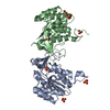


 分子量: 96.063 Da / 分子数: 8 / 由来タイプ: 合成 / 式: SO4
 分子量: 18.015 Da / 分子数: 282 / 由来タイプ: 天然 / 式: H2O
 試料調製
試料調製 / ビームライン: 3W1A / 波長: 0.95
  解析
解析