 Movie
Movie Controller


+ Open data

- Basic information
| Entry | Database: PDB / ID: 3nb2 | ||||||
|---|---|---|---|---|---|---|---|
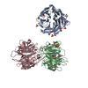
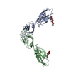
| Title | Crystal structure of E. coli O157:H7 effector protein NleL | ||||||
 Components Components | secreted effector protein | ||||||
 Keywords Keywords | LIGASE / secreted effector protein / pentapeptide / HECT domain / HECT E3 ubiquitin ligase | ||||||
| Function / homology |  Function and homology information Function and homology informationHECT-type E3 ubiquitin transferase / ubiquitin-protein transferase activity / ubiquitin protein ligase activity / host cell / protein ubiquitination / extracellular region Similarity search - Function | ||||||
| Biological species |  | ||||||
| Method |  X-RAY DIFFRACTION / SYNCHROTRON / MOLECULAR REPLACEMENT / Resolution: 2.1 Å X-RAY DIFFRACTION / SYNCHROTRON / MOLECULAR REPLACEMENT / Resolution: 2.1 Å | ||||||
 Authors Authors | Lin, D.Y. / Chen, J. | ||||||
 Citation Citation | Journal: J.Biol.Chem. / Year: 2011 Title: Biochemical and Structural Studies of a HECT-like Ubiquitin Ligase from Escherichia coli O157:H7. Authors: Lin, D.Y. / Diao, J. / Zhou, D. / Chen, J. | ||||||
| History |
|
- Structure visualization
| Structure viewer | Molecule: MolmilJmol/JSmol |
|---|
- Downloads & links
-Download
| PDBx/mmCIF format | 3nb2.cif.gz | 998.8 KB | Display | PDBx/mmCIF format |
|---|---|---|---|---|
| PDB format | pdb3nb2.ent.gz | 831.4 KB | Display | PDB format |
| PDBx/mmJSON format | 3nb2.json.gz | Tree view | PDBx/mmJSON format | |
| Others |  Other downloads Other downloads |
-Validation report
| Arichive directory | https://data.pdbj.org/pub/pdb/validation_reports/nb/3nb2ftp://data.pdbj.org/pub/pdb/validation_reports/nb/3nb2 | HTTPS FTP |
|---|
-Related structure data
| Related structure data |  3nawSC S: Starting model for refinement C: citing same article ( |
|---|---|
| Similar structure data |


-Links
 PDBj
PDBj


- Assembly
| Deposited unit | 
| ||||||||
|---|---|---|---|---|---|---|---|---|---|
| 1 | 
| ||||||||
| 2 | 
| ||||||||
| 3 | 
| ||||||||
| 4 | 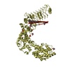
| ||||||||
| Unit cell |
|
-Components
-Protein , 1 types, 4 molecules ABCD
| #1: Protein | Mass: 69427.969 Da / Num. of mol.: 4 / Fragment: UNP residues 171-782 Source method: isolated from a genetically manipulated source Source: (gene. exp.) |
|---|
-Non-polymers , 5 types, 1150 molecules 


| #2: Chemical | ChemComp-GOL /  Mass: 92.094 Da / Num. of mol.: 29 / Source method: obtained synthetically / Formula: C3H8O3 Mass: 92.094 Da / Num. of mol.: 29 / Source method: obtained synthetically / Formula: C3H8O3#3: Chemical | ChemComp-MES /  Mass: 195.237 Da / Num. of mol.: 5 / Source method: obtained synthetically / Formula: C6H13NO4S / Comment: pH buffer*YM Mass: 195.237 Da / Num. of mol.: 5 / Source method: obtained synthetically / Formula: C6H13NO4S / Comment: pH buffer*YM#4: Chemical | ChemComp-SO4 /  Mass: 96.063 Da / Num. of mol.: 16 / Source method: obtained synthetically / Formula: SO4 Mass: 96.063 Da / Num. of mol.: 16 / Source method: obtained synthetically / Formula: SO4#5: Chemical |  Mass: 154.251 Da / Num. of mol.: 2 / Source method: obtained synthetically / Formula: C4H10O2S2 Mass: 154.251 Da / Num. of mol.: 2 / Source method: obtained synthetically / Formula: C4H10O2S2#6: Water | ChemComp-HOH / | Mass: 18.015 Da / Num. of mol.: 1098 / Source method: isolated from a natural source / Formula: H2O |
|---|
-Experimental details
-Experiment
| Experiment | Method: X-RAY DIFFRACTION / Number of used crystals: 1 |
|---|
- Sample preparation
| Crystal | Density Matthews: 2.93 Å3/Da / Density % sol: 57.96 % |
|---|---|
| Crystal grow | Temperature: 293 K / Method: vapor diffusion, sitting drop / pH: 6 Details: 0.1M Mes, 1.6M LiSO4, 16% Glyerol, VAPOR DIFFUSION, SITTING DROP, temperature 293K |
-Data collection
| Diffraction | Mean temperature: 100 K |
|---|---|
| Diffraction source | Source: SYNCHROTRON / Site: APS  / Beamline: 23-ID-D / Wavelength: 1.07223 Å / Beamline: 23-ID-D / Wavelength: 1.07223 Å |
| Detector | Type: MARMOSAIC 300 mm CCD / Detector: CCD / Date: Jul 28, 2008 / Details: Mirrors |
| Radiation | Monochromator: GRAPHITE / Protocol: SINGLE WAVELENGTH / Monochromatic (M) / Laue (L): M / Scattering type: x-ray |
| Radiation wavelength | Wavelength: 1.07223 Å / Relative weight: 1 |
| Reflection | Resolution: 2.1→50 Å / Num. all: 186206 / Num. obs: 185647 / % possible obs: 99.7 % / Observed criterion σ(F): 0 / Observed criterion σ(I): 0 / Redundancy: 3.8 % / Rsym value: 0.06 / Net I/σ(I): 22.1 |
| Reflection shell | Resolution: 2.1→2.18 Å / Redundancy: 3.8 % / Mean I/σ(I) obs: 2.6 / Rsym value: 0.547 / % possible all: 100 |
- Processing
| Software |
| |||||||||||||||||||||||||||||||||||||||||||||||||||||||||||||||||||||||||||||||||||||||||||||||||||||||||||||||||||||||||||||||||||||||||||||||||||||||||||||||||||||||||||||||||||||||||||||||||||||||||||||||||||||||||
|---|---|---|---|---|---|---|---|---|---|---|---|---|---|---|---|---|---|---|---|---|---|---|---|---|---|---|---|---|---|---|---|---|---|---|---|---|---|---|---|---|---|---|---|---|---|---|---|---|---|---|---|---|---|---|---|---|---|---|---|---|---|---|---|---|---|---|---|---|---|---|---|---|---|---|---|---|---|---|---|---|---|---|---|---|---|---|---|---|---|---|---|---|---|---|---|---|---|---|---|---|---|---|---|---|---|---|---|---|---|---|---|---|---|---|---|---|---|---|---|---|---|---|---|---|---|---|---|---|---|---|---|---|---|---|---|---|---|---|---|---|---|---|---|---|---|---|---|---|---|---|---|---|---|---|---|---|---|---|---|---|---|---|---|---|---|---|---|---|---|---|---|---|---|---|---|---|---|---|---|---|---|---|---|---|---|---|---|---|---|---|---|---|---|---|---|---|---|---|---|---|---|---|---|---|---|---|---|---|---|---|---|---|---|---|---|---|---|---|
| Refinement | Method to determine structure: MOLECULAR REPLACEMENT Starting model: PDB ENTRY 3NAW Resolution: 2.1→44.738 Å / SU ML: 0.31 / Isotropic thermal model: Anisotropic / σ(F): 1.89 / Phase error: 24.38 / Stereochemistry target values: ML
| |||||||||||||||||||||||||||||||||||||||||||||||||||||||||||||||||||||||||||||||||||||||||||||||||||||||||||||||||||||||||||||||||||||||||||||||||||||||||||||||||||||||||||||||||||||||||||||||||||||||||||||||||||||||||
| Solvent computation | Shrinkage radii: 0.9 Å / VDW probe radii: 1.11 Å / Solvent model: FLAT BULK SOLVENT MODEL / Bsol: 43.199 Å2 / ksol: 0.344 e/Å3 | |||||||||||||||||||||||||||||||||||||||||||||||||||||||||||||||||||||||||||||||||||||||||||||||||||||||||||||||||||||||||||||||||||||||||||||||||||||||||||||||||||||||||||||||||||||||||||||||||||||||||||||||||||||||||
| Displacement parameters |
| |||||||||||||||||||||||||||||||||||||||||||||||||||||||||||||||||||||||||||||||||||||||||||||||||||||||||||||||||||||||||||||||||||||||||||||||||||||||||||||||||||||||||||||||||||||||||||||||||||||||||||||||||||||||||
| Refinement step | Cycle: LAST / Resolution: 2.1→44.738 Å
| |||||||||||||||||||||||||||||||||||||||||||||||||||||||||||||||||||||||||||||||||||||||||||||||||||||||||||||||||||||||||||||||||||||||||||||||||||||||||||||||||||||||||||||||||||||||||||||||||||||||||||||||||||||||||
| Refine LS restraints |
| |||||||||||||||||||||||||||||||||||||||||||||||||||||||||||||||||||||||||||||||||||||||||||||||||||||||||||||||||||||||||||||||||||||||||||||||||||||||||||||||||||||||||||||||||||||||||||||||||||||||||||||||||||||||||
| LS refinement shell |
| |||||||||||||||||||||||||||||||||||||||||||||||||||||||||||||||||||||||||||||||||||||||||||||||||||||||||||||||||||||||||||||||||||||||||||||||||||||||||||||||||||||||||||||||||||||||||||||||||||||||||||||||||||||||||
| Refinement TLS params. | Method: refined / Origin x: -42.3929 Å / Origin y: 33.0393 Å / Origin z: 36.0663 Å
| |||||||||||||||||||||||||||||||||||||||||||||||||||||||||||||||||||||||||||||||||||||||||||||||||||||||||||||||||||||||||||||||||||||||||||||||||||||||||||||||||||||||||||||||||||||||||||||||||||||||||||||||||||||||||
| Refinement TLS group | Selection details: all |
