 Movie
Movie Controller

[English] 日本語
 Yorodumi
- PDB-3n9o: ceKDM7A from C.elegans, complex with H3K4me3 peptide, H3K9me2 pep... -
+ Open data

- Basic information
| Entry | Database: PDB / ID: 3n9o | ||||||
|---|---|---|---|---|---|---|---|
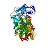
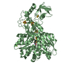
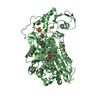
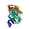
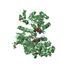
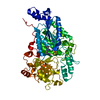
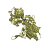
| Title | ceKDM7A from C.elegans, complex with H3K4me3 peptide, H3K9me2 peptide and NOG | ||||||
 Components Components |
| ||||||
 Keywords Keywords | OXIDOREDUCTASE / Histone / methylation / demethylase / PHD / JmjC / Fe(II) and alpha-KG (alpha-ketoglutarate)-dependent dioxygenase family | ||||||
| Function / homology |  Function and homology information Function and homology informationPKMTs methylate histone lysines / HATs acetylate histones / Negative Regulation of CDH1 Gene Transcription / Factors involved in megakaryocyte development and platelet production / Oxidative Stress Induced Senescence / RMTs methylate histone arginines / : / : / Assembly of the ORC complex at the origin of replication / RNA Polymerase I Promoter Escape ...PKMTs methylate histone lysines / HATs acetylate histones / Negative Regulation of CDH1 Gene Transcription / Factors involved in megakaryocyte development and platelet production / Oxidative Stress Induced Senescence / RMTs methylate histone arginines / : / : / Assembly of the ORC complex at the origin of replication / RNA Polymerase I Promoter Escape / RUNX1 regulates genes involved in megakaryocyte differentiation and platelet function / : / HDMs demethylate histones / Condensation of Prophase Chromosomes / histone H3K27me2/H3K27me3 demethylase activity / mitochondrial unfolded protein response / Oxidoreductases; Acting on paired donors, with incorporation or reduction of molecular oxygen; With 2-oxoglutarate as one donor, and incorporation of one atom of oxygen into each donor / histone H3K9 demethylase activity / histone demethylase activity / transcription coregulator activity / structural constituent of chromatin / nucleosome / histone binding / chromatin remodeling / protein heterodimerization activity / regulation of transcription by RNA polymerase II / DNA binding / zinc ion binding / nucleus Similarity search - Function | ||||||
| Biological species |  | ||||||
| Method |  X-RAY DIFFRACTION / SYNCHROTRON / MOLECULAR REPLACEMENT / Resolution: 2.309 Å X-RAY DIFFRACTION / SYNCHROTRON / MOLECULAR REPLACEMENT / Resolution: 2.309 Å | ||||||
 Authors Authors | Yang, Y. / Hu, L. / Wang, P. / Hou, H. / Chen, C.D. / Xu, Y. | ||||||
 Citation Citation | Journal: Cell Res. / Year: 2010 Title: Structural insights into a dual-specificity histone demethylase ceKDM7A from Caenorhabditis elegans Authors: Yang, Y. / Hu, L. / Wang, P. / Hou, H. / Lin, Y. / Liu, Y. / Li, Z. / Gong, R. / Feng, X. / Zhou, L. / Zhang, W. / Dong, Y. / Yang, H. / Lin, H. / Wang, Y. / Chen, C.D. / Xu, Y. | ||||||
| History |
|
- Structure visualization
| Structure viewer | Molecule: MolmilJmol/JSmol |
|---|
- Downloads & links
-Download
| PDBx/mmCIF format | 3n9o.cif.gz | 126.6 KB | Display | PDBx/mmCIF format |
|---|---|---|---|---|
| PDB format | pdb3n9o.ent.gz | 94.5 KB | Display | PDB format |
| PDBx/mmJSON format | 3n9o.json.gz | Tree view | PDBx/mmJSON format | |
| Others |  Other downloads Other downloads |
-Validation report
| Arichive directory | https://data.pdbj.org/pub/pdb/validation_reports/n9/3n9oftp://data.pdbj.org/pub/pdb/validation_reports/n9/3n9o | HTTPS FTP |
|---|
-Related structure data
| Related structure data | 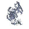 3n9lC  3n9mSC 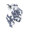 3n9nC 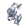 3n9pC 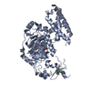 3n9qC C: citing same article ( S: Starting model for refinement |
|---|---|
| Similar structure data |


-Links
 PDBj
PDBj


- Assembly
| Deposited unit | 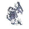
| ||||||||
|---|---|---|---|---|---|---|---|---|---|
| 1 |
| ||||||||
| Unit cell |
|
-Components
-Protein , 1 types, 1 molecules A
| #1: Protein | Mass: 61945.496 Da / Num. of mol.: 1 / Fragment: PHD domain, UNP residues 201-724 Source method: isolated from a genetically manipulated source Source: (gene. exp.)  References: UniProt: Q9GYI0, [histone H3]-dimethyl-L-lysine36 demethylase |
|---|
-Protein/peptide , 2 types, 2 molecules BC
| #2: Protein/peptide | Mass: 1607.877 Da / Num. of mol.: 1 / Fragment: JMJC domain, UNP residues 2-16 / Source method: obtained synthetically / Details: chemical synthesis, purchased from a company / Source: (synth.) |
|---|---|
| #3: Protein/peptide | Mass: 1847.152 Da / Num. of mol.: 1 / Fragment: JMJC domain, UNP residues 2-18 / Source method: obtained synthetically / Details: chemical synthesis, purchased from a company / Source: (synth.) |
-Non-polymers , 4 types, 281 molecules 


| #4: Chemical | ChemComp-FE2 /  Mass: 55.845 Da / Num. of mol.: 1 / Source method: obtained synthetically / Formula: Fe Mass: 55.845 Da / Num. of mol.: 1 / Source method: obtained synthetically / Formula: Fe | ||||
|---|---|---|---|---|---|
| #5: Chemical |  Mass: 65.409 Da / Num. of mol.: 2 / Source method: obtained synthetically / Formula: Zn Mass: 65.409 Da / Num. of mol.: 2 / Source method: obtained synthetically / Formula: Zn#6: Chemical | ChemComp-OGA / |  Mass: 147.086 Da / Num. of mol.: 1 / Source method: obtained synthetically / Formula: C4H5NO5 Mass: 147.086 Da / Num. of mol.: 1 / Source method: obtained synthetically / Formula: C4H5NO5#7: Water | ChemComp-HOH / | Mass: 18.015 Da / Num. of mol.: 277 / Source method: isolated from a natural source / Formula: H2O |
-Experimental details
-Experiment
| Experiment | Method: X-RAY DIFFRACTION / Number of used crystals: 1 |
|---|
- Sample preparation
| Crystal | Density Matthews: 2.37 Å3/Da / Density % sol: 48.11 % |
|---|---|
| Crystal grow | Temperature: 277 K / Method: vapor diffusion, hanging drop / pH: 7 Details: 16% PEG 3350, 0.2M Sodium Fluoride, 0.1M Bis-Tris or HEPES, pH 7.0, VAPOR DIFFUSION, HANGING DROP, temperature 277K |
-Data collection
| Diffraction | Mean temperature: 100 K |
|---|---|
| Diffraction source | Source: SYNCHROTRON / Site: SSRF  / Beamline: BL17U / Wavelength: 1.2828 Å / Beamline: BL17U / Wavelength: 1.2828 Å |
| Detector | Type: RAYONIX MX-225 / Detector: CCD / Date: May 6, 2009 |
| Radiation | Monochromator: SAGITALLY FOCUSED Si(111) / Protocol: SINGLE WAVELENGTH / Monochromatic (M) / Laue (L): M / Scattering type: x-ray |
| Radiation wavelength | Wavelength: 1.2828 Å / Relative weight: 1 |
| Reflection | Resolution: 2.3→50 Å / Num. obs: 27497 / % possible obs: 98.4 % / Biso Wilson estimate: 39.49 Å2 |
| Reflection shell | Resolution: 2.3→2.38 Å / % possible all: 87.3 |
- Processing
| Software |
| |||||||||||||||||||||||||||||||||||||||||||||||||||||||||||||||||||||||||||||
|---|---|---|---|---|---|---|---|---|---|---|---|---|---|---|---|---|---|---|---|---|---|---|---|---|---|---|---|---|---|---|---|---|---|---|---|---|---|---|---|---|---|---|---|---|---|---|---|---|---|---|---|---|---|---|---|---|---|---|---|---|---|---|---|---|---|---|---|---|---|---|---|---|---|---|---|---|---|---|
| Refinement | Method to determine structure: MOLECULAR REPLACEMENT Starting model: PDB ENTRY 3N9M Resolution: 2.309→44.28 Å / Occupancy max: 1 / Occupancy min: 1 / FOM work R set: 0.8261 / SU ML: 0.69 / σ(F): 1.35 / Phase error: 24.13 / Stereochemistry target values: ML
| |||||||||||||||||||||||||||||||||||||||||||||||||||||||||||||||||||||||||||||
| Solvent computation | Shrinkage radii: 0.9 Å / VDW probe radii: 1.11 Å / Solvent model: FLAT BULK SOLVENT MODEL / Bsol: 37.574 Å2 / ksol: 0.331 e/Å3 | |||||||||||||||||||||||||||||||||||||||||||||||||||||||||||||||||||||||||||||
| Displacement parameters | Biso max: 114.72 Å2 / Biso mean: 44.6602 Å2 / Biso min: 18.21 Å2
| |||||||||||||||||||||||||||||||||||||||||||||||||||||||||||||||||||||||||||||
| Refinement step | Cycle: LAST / Resolution: 2.309→44.28 Å
| |||||||||||||||||||||||||||||||||||||||||||||||||||||||||||||||||||||||||||||
| Refine LS restraints |
| |||||||||||||||||||||||||||||||||||||||||||||||||||||||||||||||||||||||||||||
| LS refinement shell |
|
