 Movie
Movie Controller

[English] 日本語
 Yorodumi
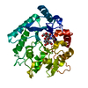
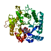
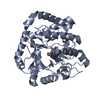
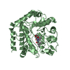
- PDB-3n11: Crystal stricture of wild-type chitinase from Bacillus cereus NCTU2 -
+ Open data

- Basic information
| Entry | Database: PDB / ID: 3n11 | ||||||
|---|---|---|---|---|---|---|---|
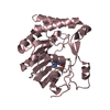
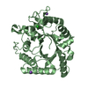
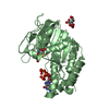
| Title | Crystal stricture of wild-type chitinase from Bacillus cereus NCTU2 | ||||||
 Components Components | Chitinase A | ||||||
 Keywords Keywords | HYDROLASE / chitinase / ChiNCTU2 / wild-type | ||||||
| Function / homology |  Function and homology information Function and homology informationendochitinase activity / chitinase / chitin binding / carbohydrate metabolic process / metal ion binding Similarity search - Function | ||||||
| Biological species |  | ||||||
| Method |  X-RAY DIFFRACTION / SYNCHROTRON / MOLECULAR REPLACEMENT / Resolution: 1.35 Å X-RAY DIFFRACTION / SYNCHROTRON / MOLECULAR REPLACEMENT / Resolution: 1.35 Å | ||||||
 Authors Authors | Hsieh, Y.-C. / Chen, C.-J. / Li, Y.-K. / Wu, Y.-J. | ||||||
 Citation Citation | Journal: J.Biol.Chem. / Year: 2010 Title: Crystal structures of bacillus cereus NCTU2 chitinase complexes with chitooligomers reveal novel substrate binding for catalysis: a chitinase without chitin-binding and insertion domains Authors: Hsieh, Y.-C. / Wu, Y.-J. / Chiang, T.-Y. / Kuo, C.-Y. / Shrestha, K.L. / Chao, C.-F. / Huang, Y.-C. / Chuankhayan, P. / Wu, W.-G. / Li, Y.-K. / Chen, C.-J. | ||||||
| History |
|
- Structure visualization
| Structure viewer | Molecule: MolmilJmol/JSmol |
|---|
- Downloads & links
-Download
| PDBx/mmCIF format | 3n11.cif.gz | 82.6 KB | Display | PDBx/mmCIF format |
|---|---|---|---|---|
| PDB format | pdb3n11.ent.gz | 60.1 KB | Display | PDB format |
| PDBx/mmJSON format | 3n11.json.gz | Tree view | PDBx/mmJSON format | |
| Others |  Other downloads Other downloads |
-Validation report
| Arichive directory | https://data.pdbj.org/pub/pdb/validation_reports/n1/3n11ftp://data.pdbj.org/pub/pdb/validation_reports/n1/3n11 | HTTPS FTP |
|---|
-Related structure data
| Related structure data | 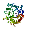 3n12SC 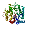 3n13C  3n15C  3n17C 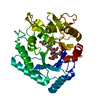 3n18C 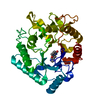 3n1aC S: Starting model for refinement C: citing same article ( |
|---|---|
| Similar structure data |


-Links
 PDBj
PDBj- Assembly
| Deposited unit | 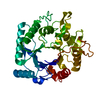
| ||||||||
|---|---|---|---|---|---|---|---|---|---|
| 1 |
| ||||||||
| Unit cell |
|
-Components
| #1: Protein | Mass: 36267.625 Da / Num. of mol.: 1 Source method: isolated from a genetically manipulated source Source: (gene. exp.) |
|---|---|
| #2: Water | ChemComp-HOH /  Mass: 18.015 Da / Num. of mol.: 342 / Source method: isolated from a natural source / Formula: H2O Mass: 18.015 Da / Num. of mol.: 342 / Source method: isolated from a natural source / Formula: H2O |
| Sequence details | THERE IS CONFLICT BETWEEN THE REPORTED SEQUENCE AND DATABASE REFERENCE SEQUENCE. ACCORDING TO THE ...THERE IS CONFLICT BETWEEN THE REPORTED SEQUENCE AND DATABASE REFERENCE SEQUENCE. ACCORDING TO THE ELECTRON DENSITY THE POSITION 277 IS OBVIOUSLY VAL THAN ALA. |
-Experimental details
-Experiment
| Experiment | Method: X-RAY DIFFRACTION / Number of used crystals: 2 |
|---|
- Sample preparation
| Crystal | Density Matthews: 1.95 Å3/Da / Density % sol: 36.88 % |
|---|---|
| Crystal grow | Temperature: 291 K / Method: vapor diffusion, hanging drop / pH: 6 Details: 50mM potassium phosphate monobasic, 20%(w/v) PEG 8000, pH 6.0, VAPOR DIFFUSION, HANGING DROP, temperature 291K |
-Data collection
| Diffraction | Mean temperature: 110 K |
|---|---|
| Diffraction source | Source: SYNCHROTRON / Site: NSRRC  / Beamline: BL13B1 / Wavelength: 1 Å / Beamline: BL13B1 / Wavelength: 1 Å |
| Detector | Type: ADSC QUANTUM 315 / Detector: CCD / Date: Jan 12, 2008 |
| Radiation | Protocol: SINGLE WAVELENGTH / Monochromatic (M) / Laue (L): M / Scattering type: x-ray |
| Radiation wavelength | Wavelength: 1 Å / Relative weight: 1 |
| Reflection | Resolution: 1.35→30 Å / Num. all: 62982 / Num. obs: 62227 / % possible obs: 98.9 % / Redundancy: 5.9 % / Rmerge(I) obs: 0.049 / Rsym value: 0.049 / Net I/σ(I): 34.7 |
| Reflection shell | Resolution: 1.35→1.4 Å / Redundancy: 5.5 % / Rmerge(I) obs: 0.28 / Mean I/σ(I) obs: 6.2 / Num. unique all: 5979 / Rsym value: 0.239 / % possible all: 96.5 |
- Processing
| Software |
| |||||||||||||||||||||||||||||||||||||||||||||||||||||||||||||||||
|---|---|---|---|---|---|---|---|---|---|---|---|---|---|---|---|---|---|---|---|---|---|---|---|---|---|---|---|---|---|---|---|---|---|---|---|---|---|---|---|---|---|---|---|---|---|---|---|---|---|---|---|---|---|---|---|---|---|---|---|---|---|---|---|---|---|---|
| Refinement | Method to determine structure: MOLECULAR REPLACEMENT Starting model: PDB ENTRY 3N12 Resolution: 1.35→20 Å / Cor.coef. Fo:Fc: 0.956 / Cor.coef. Fo:Fc free: 0.95 / SU B: 0.995 / SU ML: 0.042 / Cross valid method: THROUGHOUT / ESU R Free: 0.065 / Stereochemistry target values: MAXIMUM LIKELIHOOD / Details: HYDROGENS HAVE BEEN ADDED IN THE RIDING POSITIONS
| |||||||||||||||||||||||||||||||||||||||||||||||||||||||||||||||||
| Solvent computation | Ion probe radii: 0.8 Å / Shrinkage radii: 0.8 Å / VDW probe radii: 1.2 Å / Solvent model: MASK | |||||||||||||||||||||||||||||||||||||||||||||||||||||||||||||||||
| Displacement parameters | Biso mean: 13.532 Å2
| |||||||||||||||||||||||||||||||||||||||||||||||||||||||||||||||||
| Refinement step | Cycle: LAST / Resolution: 1.35→20 Å
| |||||||||||||||||||||||||||||||||||||||||||||||||||||||||||||||||
| Refine LS restraints |
| |||||||||||||||||||||||||||||||||||||||||||||||||||||||||||||||||
| LS refinement shell | Resolution: 1.35→1.385 Å / Total num. of bins used: 20
|
