 Movie
Movie Controller

[English] 日本語
 Yorodumi
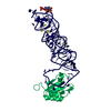
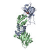
- PDB-3muv: Crystal Structure of the G20A/C92U mutant c-di-GMP riboswith boun... -
+ Open data

- Basic information
| Entry | Database: PDB / ID: 3muv | ||||||
|---|---|---|---|---|---|---|---|
| Title | Crystal Structure of the G20A/C92U mutant c-di-GMP riboswith bound to c-di-AMP | ||||||
 Components Components |
| ||||||
 Keywords Keywords | RNA binding PROTEIN/rna / RNA / riboswitch / c-di-AMP / c-di-GMP / RNA binding PROTEIN-rna complex | ||||||
| Function / homology |  Function and homology information Function and homology informationU1 snRNP binding / U1 snRNP / U1 snRNA binding / U4/U6 x U5 tri-snRNP complex / mRNA Splicing - Major Pathway / spliceosomal complex / mRNA splicing, via spliceosome / DNA binding / RNA binding / nucleoplasm ...U1 snRNP binding / U1 snRNP / U1 snRNA binding / U4/U6 x U5 tri-snRNP complex / mRNA Splicing - Major Pathway / spliceosomal complex / mRNA splicing, via spliceosome / DNA binding / RNA binding / nucleoplasm / identical protein binding / nucleus Similarity search - Function | ||||||
| Biological species |  Homo sapiens (human) Homo sapiens (human) | ||||||
| Method |  X-RAY DIFFRACTION / SYNCHROTRON / MOLECULAR REPLACEMENT / Resolution: 3.2 Å X-RAY DIFFRACTION / SYNCHROTRON / MOLECULAR REPLACEMENT / Resolution: 3.2 Å | ||||||
 Authors Authors | Strobel, S.A. / Smith, K.D. | ||||||
 Citation Citation | Journal: Biochemistry / Year: 2010 Title: Structural and biochemical determinants of ligand binding by the c-di-GMP riboswitch . Authors: Smith, K.D. / Lipchock, S.V. / Livingston, A.L. / Shanahan, C.A. / Strobel, S.A. | ||||||
| History |
|
- Structure visualization
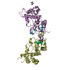
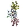
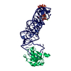
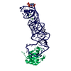
| Structure viewer | Molecule: MolmilJmol/JSmol |
|---|
- Downloads & links
-Download
| PDBx/mmCIF format | 3muv.cif.gz | 154.4 KB | Display | PDBx/mmCIF format |
|---|---|---|---|---|
| PDB format | pdb3muv.ent.gz | 117.8 KB | Display | PDB format |
| PDBx/mmJSON format | 3muv.json.gz | Tree view | PDBx/mmJSON format | |
| Others |  Other downloads Other downloads |
-Validation report
| Arichive directory | https://data.pdbj.org/pub/pdb/validation_reports/mu/3muvftp://data.pdbj.org/pub/pdb/validation_reports/mu/3muv | HTTPS FTP |
|---|
-Related structure data
| Related structure data | 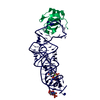 3mumC  3murC  3mutC 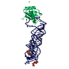 3mxhC  3irwS C: citing same article ( S: Starting model for refinement |
|---|---|
| Similar structure data |


-Links
 PDBj
PDBj


- Assembly
| Deposited unit | 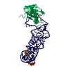
| ||||||||
|---|---|---|---|---|---|---|---|---|---|
| 1 |
| ||||||||
| Unit cell |
|
-Components
| #1: Protein | Mass: 11340.315 Da / Num. of mol.: 1 / Fragment: UNP residues 1-98 / Mutation: Y31H, Q36R Source method: isolated from a genetically manipulated source Source: (gene. exp.) Homo sapiens (human) / Gene: SNRPA / Plasmid: pET11 / Production host:  |
|---|---|
| #2: RNA chain | Mass: 29927.758 Da / Num. of mol.: 1 / Source method: obtained synthetically / Details: in vitro transcribed from linear DNA |
| #3: Chemical | ChemComp-2BA / (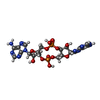  Mass: 658.412 Da / Num. of mol.: 1 / Source method: obtained synthetically / Formula: C20H24N10O12P2 Mass: 658.412 Da / Num. of mol.: 1 / Source method: obtained synthetically / Formula: C20H24N10O12P2 |
| #4: Chemical | ChemComp-MG /   Mass: 24.305 Da / Num. of mol.: 1 / Source method: obtained synthetically / Formula: Mg Mass: 24.305 Da / Num. of mol.: 1 / Source method: obtained synthetically / Formula: Mg |
| #5: Water | ChemComp-HOH /  Mass: 18.015 Da / Num. of mol.: 16 / Source method: isolated from a natural source / Formula: H2O Mass: 18.015 Da / Num. of mol.: 16 / Source method: isolated from a natural source / Formula: H2O |
-Experimental details
-Experiment
| Experiment | Method: X-RAY DIFFRACTION / Number of used crystals: 1 |
|---|
- Sample preparation
| Crystal | Density Matthews: 2.05 Å3/Da / Density % sol: 39.93 % |
|---|---|
| Crystal grow | Temperature: 298 K / pH: 6 Details: 25% PEG 550 MME, 5 mM MgSO4, 50 mM MES, pH 6.0, 300 mM NaCl, VAPOR DIFFUSION, HANGING DROP, temperature 298K |
-Data collection
| Diffraction | Mean temperature: 100 K |
|---|---|
| Diffraction source | Source: SYNCHROTRON / Site: NSLS  / Beamline: X25 / Wavelength: 1.1 / Beamline: X25 / Wavelength: 1.1 |
| Detector | Type: ADSC QUANTUM 315 / Detector: CCD / Date: Nov 19, 2009 / Details: PT-COATED MIRROR |
| Radiation | Monochromator: SI-111 DOUBLE CRYSTAL / Protocol: SINGLE WAVELENGTH / Monochromatic (M) / Laue (L): M / Scattering type: x-ray |
| Radiation wavelength | Wavelength: 1.1 Å / Relative weight: 1 |
| Reflection | Resolution: 3.2→80 Å / Num. obs: 4829 / % possible obs: 90 % / Redundancy: 3.2 % / Rmerge(I) obs: 0.197 / Net I/σ(I): 5.6 |
| Reflection shell | Resolution: 3.2→3.3 Å / Redundancy: 2.2 % / Rmerge(I) obs: 0.333 / Mean I/σ(I) obs: 2.2 / % possible all: 63.6 |
- Processing
| Software |
| ||||||||||||||||||||||||||||||||||||||||||||||||||||||||||||||||||||||||||||||||||||||||||||||||||||||
|---|---|---|---|---|---|---|---|---|---|---|---|---|---|---|---|---|---|---|---|---|---|---|---|---|---|---|---|---|---|---|---|---|---|---|---|---|---|---|---|---|---|---|---|---|---|---|---|---|---|---|---|---|---|---|---|---|---|---|---|---|---|---|---|---|---|---|---|---|---|---|---|---|---|---|---|---|---|---|---|---|---|---|---|---|---|---|---|---|---|---|---|---|---|---|---|---|---|---|---|---|---|---|---|
| Refinement | Method to determine structure: MOLECULAR REPLACEMENT Starting model: PDB ENTRY 3IRW Resolution: 3.2→48.65 Å / Cross valid method: THROUGHOUT / Stereochemistry target values: MAXIMUM LIKELIHOOD
| ||||||||||||||||||||||||||||||||||||||||||||||||||||||||||||||||||||||||||||||||||||||||||||||||||||||
| Displacement parameters | Biso mean: 41.09 Å2
| ||||||||||||||||||||||||||||||||||||||||||||||||||||||||||||||||||||||||||||||||||||||||||||||||||||||
| Refinement step | Cycle: LAST / Resolution: 3.2→48.65 Å
| ||||||||||||||||||||||||||||||||||||||||||||||||||||||||||||||||||||||||||||||||||||||||||||||||||||||
| Refine LS restraints |
| ||||||||||||||||||||||||||||||||||||||||||||||||||||||||||||||||||||||||||||||||||||||||||||||||||||||
| LS refinement shell | Resolution: 3.2→3.28 Å
| ||||||||||||||||||||||||||||||||||||||||||||||||||||||||||||||||||||||||||||||||||||||||||||||||||||||
| Refinement TLS params. | Method: refined / Refine-ID: X-RAY DIFFRACTION
| ||||||||||||||||||||||||||||||||||||||||||||||||||||||||||||||||||||||||||||||||||||||||||||||||||||||
| Refinement TLS group |
|
