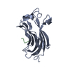
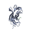
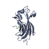
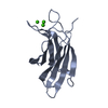
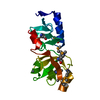
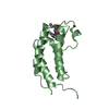
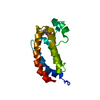
- PDB-3mqs: Crystal Structure of the USP7:Hdm2(PSTS) complex -
+
データを開く
IDまたはキーワード:
読み込み中...
-
基本情報
登録情報
データベース: PDB / ID: 3mqs
タイトル
Crystal Structure of the USP7:Hdm2(PSTS) complex
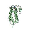
要素
Hdm2 peptide
Ubiquitin carboxyl-terminal hydrolase 7
キーワード
HYDROLASE / USP7
機能・相同性
機能・相同性情報
regulation of telomere capping / regulation of establishment of protein localization to telomere / monoubiquitinated protein deubiquitination / cellular response to vitamin B1 / response to formaldehyde / response to water-immersion restraint stress / response to ether / regulation of retrograde transport, endosome to Golgi / traversing start control point of mitotic cell cycle / atrial septum development ...regulation of telomere capping / regulation of establishment of protein localization to telomere / monoubiquitinated protein deubiquitination / cellular response to vitamin B1 / response to formaldehyde / response to water-immersion restraint stress / response to ether / regulation of retrograde transport, endosome to Golgi / traversing start control point of mitotic cell cycle / atrial septum development / regulation of protein catabolic process at postsynapse, modulating synaptic transmission / fibroblast activation / deubiquitinase activity / Trafficking of AMPA receptors / receptor serine/threonine kinase binding / DNA alkylation repair / regulation of DNA-binding transcription factor activity / peroxisome proliferator activated receptor binding / negative regulation of intrinsic apoptotic signaling pathway by p53 class mediator / positive regulation of vascular associated smooth muscle cell migration / negative regulation of protein processing / K48-linked deubiquitinase activity / SUMO transferase activity / response to steroid hormone / NEDD8 ligase activity / AKT phosphorylates targets in the cytosol / response to iron ion / atrioventricular valve morphogenesis / symbiont-mediated disruption of host cell PML body / endocardial cushion morphogenesis / cellular response to peptide hormone stimulus / ventricular septum development / negative regulation of NF-kappaB transcription factor activity / positive regulation of muscle cell differentiation / negative regulation of gene expression via chromosomal CpG island methylation / cardiac septum morphogenesis / SUMOylation of ubiquitinylation proteins / regulation of postsynaptic neurotransmitter receptor internalization / blood vessel development / ligase activity / cellular response to alkaloid / Constitutive Signaling by AKT1 E17K in Cancer / regulation of protein catabolic process / negative regulation of signal transduction by p53 class mediator / negative regulation of DNA damage response, signal transduction by p53 class mediator / SUMOylation of transcription factors / response to magnesium ion / cellular response to UV-C / protein sumoylation / cellular response to actinomycin D / blood vessel remodeling / protein deubiquitination / cellular response to estrogen stimulus / protein localization to nucleus / negative regulation of gluconeogenesis / ribonucleoprotein complex binding / protein autoubiquitination / positive regulation of vascular associated smooth muscle cell proliferation / negative regulation of TORC1 signaling / NPAS4 regulates expression of target genes / transcription-coupled nucleotide-excision repair / negative regulation of proteasomal ubiquitin-dependent protein catabolic process / transcription repressor complex / positive regulation of mitotic cell cycle / regulation of heart rate / Regulation of PTEN localization / proteolysis involved in protein catabolic process / Synthesis of active ubiquitin: roles of E1 and E2 enzymes / regulation of signal transduction by p53 class mediator / positive regulation of protein export from nucleus / ubiquitin binding / response to cocaine / DNA damage response, signal transduction by p53 class mediator / Stabilization of p53 / establishment of protein localization / Regulation of RUNX3 expression and activity / cellular response to gamma radiation / regulation of circadian rhythm / PML body / Oncogene Induced Senescence / protein destabilization / RING-type E3 ubiquitin transferase / Regulation of TP53 Activity through Methylation / regulation of protein stability / cellular response to growth factor stimulus / response to toxic substance / Transcription-Coupled Nucleotide Excision Repair (TC-NER) / Formation of TC-NER Pre-Incision Complex / centriolar satellite / cellular response to hydrogen peroxide / protein polyubiquitination / Dual incision in TC-NER / Gap-filling DNA repair synthesis and ligation in TC-NER / ubiquitin-protein transferase activity / disordered domain specific binding / p53 binding / endocytic vesicle membrane / ubiquitin protein ligase activity / Signaling by ALK fusions and activated point mutants / rhythmic process 類似検索 - 分子機能
MATH domain / Apoptosis, Tumor Necrosis Factor Receptor Associated Protein 2; Chain A / Apoptosis, Tumor Necrosis Factor Receptor Associated Protein 2; Chain A / Ubiquitin carboxyl-terminal hydrolase 7, ICP0-binding domain / ICP0-binding domain of Ubiquitin-specific protease 7 / Ubiquitin carboxyl-terminal hydrolase, C-terminal / Ubiquitin-specific protease C-terminal / MATH domain / E3 ubiquitin-protein ligase Mdm2 / MDM2, modified RING finger, HC subclass ...MATH domain / Apoptosis, Tumor Necrosis Factor Receptor Associated Protein 2; Chain A / Apoptosis, Tumor Necrosis Factor Receptor Associated Protein 2; Chain A / Ubiquitin carboxyl-terminal hydrolase 7, ICP0-binding domain / ICP0-binding domain of Ubiquitin-specific protease 7 / Ubiquitin carboxyl-terminal hydrolase, C-terminal / Ubiquitin-specific protease C-terminal / MATH domain / E3 ubiquitin-protein ligase Mdm2 / MDM2, modified RING finger, HC subclass / : / MATH/TRAF domain / MATH/TRAF domain profile. / meprin and TRAF homology / p53 negative regulator Mdm2/Mdm4 / TRAF-like / SWIB/MDM2 domain / SWIB/MDM2 domain / SWIB/MDM2 domain profile. / SWIB/MDM2 domain superfamily / Ubiquitin specific protease (USP) domain signature 2. / Ubiquitin specific protease (USP) domain signature 1. / Ubiquitin specific protease, conserved site / Peptidase C19, ubiquitin carboxyl-terminal hydrolase / Ubiquitin carboxyl-terminal hydrolase / Zn-finger in Ran binding protein and others / Ubiquitin specific protease domain / Ubiquitin specific protease (USP) domain profile. / Zinc finger, C3HC4 type (RING finger) / Zinc finger RanBP2 type profile. / Zinc finger, RanBP2-type superfamily / Zinc finger RanBP2-type signature. / Zinc finger, RanBP2-type / Papain-like cysteine peptidase superfamily / Zinc finger RING-type profile. / Zinc finger, RING-type / Zinc finger, RING/FYVE/PHD-type / Sandwich / Mainly Beta 類似検索 - ドメイン・相同性
 ムービー
ムービー コントローラー


 データを開く
 基本情報
基本情報 要素
要素 キーワード
キーワード 機能・相同性情報
 Homo sapiens (ヒト)
 X線回折 /
  データ登録者
 引用
引用 構造の表示
構造の表示 ダウンロードとリンク
 その他のダウンロード


 PDBj
PDBj


 集合体
集合体

 分子量: 18.015 Da / 分子数: 102 / 由来タイプ: 天然 / 式: H2O
 試料調製
試料調製 解析
解析