 Movie
Movie Controller

[English] 日本語
 Yorodumi
- PDB-3mnd: Crystallographic analysis of the cystosolic cu/zn Superoxide dism... -
+ Open data

- Basic information
| Entry | Database: PDB / ID: 3mnd | ||||||
|---|---|---|---|---|---|---|---|
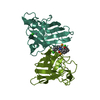
| Title | Crystallographic analysis of the cystosolic cu/zn Superoxide dismutase from taenia solium | ||||||
 Components Components | Superoxide dismutase [Cu-Zn] | ||||||
 Keywords Keywords | OXIDOREDUCTASE / CU/ZN-SUPEROXIDE DISMUTASE / METAL-MEDIATED SELF-ASSEMBLY | ||||||
| Function / homology |  Function and homology information Function and homology informationsuperoxide dismutase / superoxide dismutase activity / copper ion binding / identical protein binding Similarity search - Function | ||||||
| Biological species |  Taenia solium (pork tapeworm) Taenia solium (pork tapeworm) | ||||||
| Method |  X-RAY DIFFRACTION / SYNCHROTRON / MOLECULAR REPLACEMENT / Resolution: 2.2 Å X-RAY DIFFRACTION / SYNCHROTRON / MOLECULAR REPLACEMENT / Resolution: 2.2 Å | ||||||
 Authors Authors | Hernandez-Santoyo, A. / Rodriguez-Romero, A. | ||||||
 Citation Citation | Journal: Febs J. / Year: 2011 Title: Crystal structure of Cu / Zn superoxide dismutase from Taenia solium reveals metal-mediated self-assembly Authors: Hernandez-Santoyo, A. / Landa, A. / Gonzalez-Mondragon, E. / Pedraza-Escalona, M. / Parra-Unda, R. / Rodriguez-Romero, A. #1: Journal: Int.J.Parasitol. / Year: 2002 Title: Cloning, production and characterisation of a recombinant Cu/Zn superoxide dismutase from Taenia solium. Authors: Castellanos-Gonzalez, A. / Jimenez, L. / Landa, A. | ||||||
| History |
|
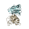
- Structure visualization
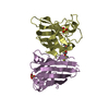
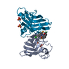
| Structure viewer | Molecule: MolmilJmol/JSmol |
|---|
- Downloads & links
-Download
| PDBx/mmCIF format | 3mnd.cif.gz | 70.1 KB | Display | PDBx/mmCIF format |
|---|---|---|---|---|
| PDB format | pdb3mnd.ent.gz | 52 KB | Display | PDB format |
| PDBx/mmJSON format | 3mnd.json.gz | Tree view | PDBx/mmJSON format | |
| Others |  Other downloads Other downloads |
-Validation report
| Arichive directory | https://data.pdbj.org/pub/pdb/validation_reports/mn/3mndftp://data.pdbj.org/pub/pdb/validation_reports/mn/3mnd | HTTPS FTP |
|---|
-Related structure data
| Related structure data | 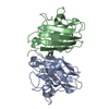 1to4S S: Starting model for refinement |
|---|---|
| Similar structure data |


-Links
 PDBj
PDBj
- Assembly
| Deposited unit | 
| ||||||||
|---|---|---|---|---|---|---|---|---|---|
| 1 |
| ||||||||
| Unit cell |
|
-Components
| #1: Protein | Mass: 15613.516 Da / Num. of mol.: 2 Source method: isolated from a genetically manipulated source Details: EXPRESSION_SYSTEM_CELLULAR_LOCATION: CYTOPLASM / Source: (gene. exp.) Taenia solium (pork tapeworm) / Strain: MEXICAN STRAIN / Plasmid: PRSETB / Production host:  #2: Chemical | ChemComp-ZN /   Mass: 65.409 Da / Num. of mol.: 5 / Source method: obtained synthetically / Formula: Zn Mass: 65.409 Da / Num. of mol.: 5 / Source method: obtained synthetically / Formula: Zn#3: Chemical |   Mass: 63.546 Da / Num. of mol.: 2 / Source method: obtained synthetically / Formula: Cu Mass: 63.546 Da / Num. of mol.: 2 / Source method: obtained synthetically / Formula: Cu#4: Chemical |   Mass: 92.094 Da / Num. of mol.: 2 / Source method: obtained synthetically / Formula: C3H8O3 Mass: 92.094 Da / Num. of mol.: 2 / Source method: obtained synthetically / Formula: C3H8O3#5: Water | ChemComp-HOH / |  Mass: 18.015 Da / Num. of mol.: 109 / Source method: isolated from a natural source / Formula: H2O Mass: 18.015 Da / Num. of mol.: 109 / Source method: isolated from a natural source / Formula: H2OHas protein modification | Y | |
|---|
-Experimental details
-Experiment
| Experiment | Method: X-RAY DIFFRACTION / Number of used crystals: 1 |
|---|
- Sample preparation
| Crystal | Density Matthews: 2.08 Å3/Da / Density % sol: 42.24 % |
|---|---|
| Crystal grow | Temperature: 298 K / pH: 8.5 Details: 20% w/v polyethylene glycol monomethyl ether 2,000, in hexahydrate 0.1 M Tris buffer pH 8.5 and 0.01 M Nickel (II) chloridE., VAPOR DIFFUSION, HANGING DROP, temperature 298K |
-Data collection
| Diffraction | Mean temperature: 100 K |
|---|---|
| Diffraction source | Source: SYNCHROTRON / Site: NSLS  / Beamline: X12B / Wavelength: 0.9795 / Beamline: X12B / Wavelength: 0.9795 |
| Detector | Type: ADSC QUANTUM 4 / Detector: CCD / Date: Apr 14, 2005 / Details: MIRRORS |
| Radiation | Monochromator: GRAPHITE / Protocol: SINGLE WAVELENGTH / Monochromatic (M) / Laue (L): M / Scattering type: x-ray |
| Radiation wavelength | Wavelength: 0.9795 Å / Relative weight: 1 |
| Reflection | Resolution: 2.2→39.64 Å / Num. obs: 13829 / % possible obs: 97.7 % / Redundancy: 3.2 % / Biso Wilson estimate: 20.94 Å2 / Rmerge(I) obs: 0.116 / Net I/σ(I): 7 |
| Reflection shell | Highest resolution: 2.2 Å / Redundancy: 3 % / Rmerge(I) obs: 0.356 / Mean I/σ(I) obs: 2.8 / % possible all: 98.8 |
- Processing
| Software |
| |||||||||||||||||||||||||||||||||||||||||||||||||||||||||||||||||||||||||||||
|---|---|---|---|---|---|---|---|---|---|---|---|---|---|---|---|---|---|---|---|---|---|---|---|---|---|---|---|---|---|---|---|---|---|---|---|---|---|---|---|---|---|---|---|---|---|---|---|---|---|---|---|---|---|---|---|---|---|---|---|---|---|---|---|---|---|---|---|---|---|---|---|---|---|---|---|---|---|---|
| Refinement | Method to determine structure: MOLECULAR REPLACEMENT Starting model: 1TO4 Resolution: 2.2→39.64 Å / SU ML: 0.28 / Isotropic thermal model: Isotropic / σ(F): 0.09 / Phase error: 23.23 / Stereochemistry target values: ML
| |||||||||||||||||||||||||||||||||||||||||||||||||||||||||||||||||||||||||||||
| Solvent computation | Shrinkage radii: 0.83 Å / VDW probe radii: 1.1 Å / Solvent model: FLAT BULK SOLVENT MODEL / Bsol: 33.96 Å2 / ksol: 0.38 e/Å3 | |||||||||||||||||||||||||||||||||||||||||||||||||||||||||||||||||||||||||||||
| Refinement step | Cycle: LAST / Resolution: 2.2→39.64 Å
| |||||||||||||||||||||||||||||||||||||||||||||||||||||||||||||||||||||||||||||
| Refine LS restraints |
| |||||||||||||||||||||||||||||||||||||||||||||||||||||||||||||||||||||||||||||
| LS refinement shell |
|
